
Page 58 - Feline diagnostic imaging
P. 58
54 5 Diagnostic Imaging of Diseases of the Skull
(a) (b)
(c)
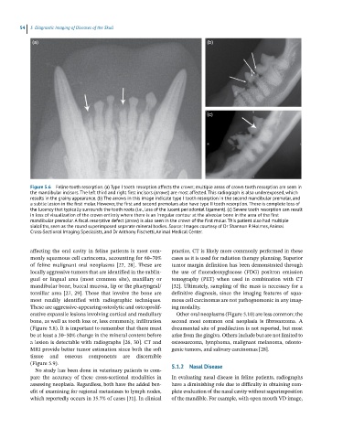
Figure 5.6 Feline tooth resorption. (a) Type I tooth resorption affects the crown; multiple areas of crown tooth resorption are seen in
the mandibular incisors. The left third and right first incisors (arrows) are most affected. This radiograph is also underexposed, which
results in the grainy appearance. (b) The arrows in this image indicate type I tooth resorption in the second mandibular premolar, and
a subtle lesion in the first molar. However, the first and second premolars also have type II tooth resorption. There is complete loss of
the lucency that typically surrounds the tooth roots (i.e., loss of the lucent periodontal ligament). (c) Severe tooth resorption can result
in loss of visualization of the crown entirely where there is an irregular contour at the alveolar bone in the area of the first
mandibular premolar. A focal resorptive defect (arrow) is also seen in the crown of the first molar. This patient also had multiple
sialoliths, seen as the round superimposed separate mineral bodies. Source: Images courtesy of Dr Shannon P. Holmes, Animal
Cross‐Sectional Imaging Specialists, and Dr Anthony Fischetti, Animal Medical Center.
affecting the oral cavity in feline patients is most com- practice, CT is likely more commonly performed in these
monly squamous cell carincoma, accounting for 60–70% cases as it is used for radiation therapy planning. Superior
of feline malignant oral neoplasms [27, 28]. These are tumor margin definition has been demonstrated through
locally aggressive tumors that are identified in the sublin- the use of fluorodeoxyglucose (FDG) positron emission
gual or lingual area (most common site), maxillary or tomography (PET) when used in combination with CT
mandibular bone, buccal mucosa, lip or the pharyngeal/ [32]. Ultimately, sampling of the mass is necessary for a
tonsillar area [27, 29]. Those that involve the bone are definitive diagnosis, since the imaging features of squa-
most readily identified with radiographic techniques. mous cell carcinomas are not pathognomonic in any imag-
These are aggressive‐appearing osteolytic and osteoprolif- ing modality.
erative expansile lesions involving cortical and medullary Other oral neoplasms (Figure 5.10) are less common; the
bone, as well as tooth loss or, less commonly, infiltration second most common oral neoplasia is fibrosarcoma. A
(Figure 5.8). It is important to remember that there must documented site of predilection is not reported, but most
be at least a 30–50% change in the mineral content before arise from the gingiva. Others include but are not limited to
a lesion is detectable with radiographs [26, 30]. CT and osteosarcoma, lymphoma, malignant melanoma, odonto-
MRI provide better tumor estimation since both the soft genic tumors, and salivary carcinomas [28].
tissue and osseous components are discernible
(Figure 5.9). 5.1.2 Nasal Disease
No study has been done in veterinary patients to com-
pare the accuracy of these cross‐sectional modalities in In evaluating nasal disease in feline patients, radiographs
assessing neoplasia. Regardless, both have the added ben- have a diminishing role due to difficulty in obtaining com-
efit of examining for regional metastases to lymph nodes, plete evaluation of the nasal cavity without superimposition
which reportedly occurs in 35.7% of cases [31]. In clinical of the mandible. For example, with open mouth VD image,