
Page 55 - Feline diagnostic imaging
P. 55
5.1 Diseases of the eline Skull 51
(a) (b)
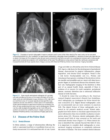
Figure 5.2 Examples of special radiographic images to evaluate specific areas of the feline skull. (a) The nasal cavity can be evaluated
without obstruction from the mandible in an open‐mouth ventrodorsal radiograph. General anesthesia is necessary to maximally open the jaw
to obtain this image. (b) The tympanic bullae (arrows) can be assessed for symmetry and with less superimposed anatomy with a rostrocaudal
open‐mouth ventrodorsal projection with ventral flexion of the neck. The left bulla is normal and air‐filled; the right bulla is abnormal with
increased luminal soft tissue opacity. Source: Images courtesy of Dr Shannon P. Holmes, Animal Cross‐Sectional Imaging Specialists.
of age and older are affected by some form of dental disease
[16]. Age is a risk factor for the development of periodontal
disease, characterized by gingival inflammation, plaque
deposition, and alveolar bone resorption. Breed is also
a risk factor; brachycephalic cats (i.e., Persian and
Himalayan) have greater crowding of the dentition within
the maxilla and mandible and as a result will often have a
greater degree of periodontal disease than domestic short-
hair cats. Radiographic imaging of the dentition is often
part of an annual health check, especially if there is
evidence of or concern for tooth resorption, periodontal
disease, anatomic distortion due to abnormal infiltrative
disease or tooth fractures.
Figure 5.3 Open‐mouth ventrodorsal radiograph of a geriatric It should be noted that according to the American
cat under anesthesia. Portions of the nasal turbinates of the left Animal Hospital Association’s Dental Care Guidelines,
nasal cavity are well delineated by gas, whereas on the right there
are multifocal areas of increased opacity (arrows) and the nasal skull radiographs are inadequate for feline dental dis -
turbinates are less well defined in these areas. A CT examination ease evaluation [17]. Digital dental radiographic units
of this patient showed no nasal abnormalities demonstrating the are recommended and are more common in practices
effect of poor positioning that could lead to misdiagnosis. Patients [18]. The digital format of these radiographs can be
like this, with multiple missing teeth, can be more difficult to
position and assess for symmetry. Source: Images courtesy of Dr stored in the patient’s medical record. Dental radio -
Shannon P. Holmes, Animal Cross‐Sectional Imaging Specialists. graphs have been shown to elucidate approximately 42%
more information than was available on physical exami-
5.1 Diseases of the Feline Skull nation alone [19]. However, dental radiographs offer a
focused small field of view centered on the teeth and
5.1.1 Dental Disease associated alveolar bone (Figure 5.5). When dental dis-
ease is identified that extends beyond this field of view,
In feline patients, a range of abnormalities affecting the it is important to consider skull radiographs or cross‐
teeth or associated maxillary or mandibular bone are rou- sectional imaging techniques to completely delineate
tinely encountered. It is estimated that 68% of cats 3 years the extent of disease. The most common dental diseases