
Page 60 - Feline diagnostic imaging
P. 60
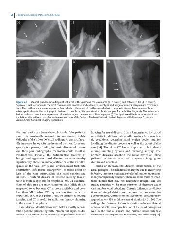
56  5  Diagnostic Imaging of Diseases of the Skull

            (a)                                  (b)                       (c)


            (d)                                   (e)                    (f)


            Figure 5.8  Intraoral mandibular radiographs of a cat with squamous cell carcinoma (a–c, arrows) and osteomyelitis (d–e, arrows).
            Squamous cell carcinoma is the most common oral neoplasm and extensive osteolysis and irregular mineral margins are commonly
            seen. The teeth in some areas appear to float, which is the result of teeth embedded with neoplastic tissue. Because mandibular
            osteomyelitis has similar radiographic features to neoplasia, it is important to obtain samples for definitive diagnosis. The extent of a
            lesion, such as a mandibular squamous cell carcinoma, can be seen in skull radiographs (f). The right mandible is more ventral than
            the left on this oblique view. Source: Images courtesy of Dr Anthony Fischetti, Animal Medical Center, and Dr Shannon P. Holmes,
            Animal Cross‐Sectional Imaging Specialists.


            the nasal cavity can be evaluated but only if the patient’s   imaging for nasal disease. It has demonstrated increased
            mouth  is  maximally  opened.  As  mentioned,  subtle   sensitivity for differentiating inflammatory from neoplas-
              obliquity of the VD or DV skull radiograph can artifactu-  tic  conditions,  detecting  nasal  foreign  bodies  and  for
            ally increase the opacity in the nasal cavities. Increased   localizing the disease process as well as the extent of dis-
            opacity is a primary finding in most feline nasal disease   ease [34]. Therefore, CT has an important role in deter-
            and  thus  poor  radiographic  technique  could  result  in   mining  sampling  options  and  planning  surgery.  The
            misdiagnosis.  Finally,  the  radiographic  features  of   primary  diseases  affecting  the  nasal  cavity  of  feline
            benign  and  aggressive  nasal  disease  processes  overlap   patients that are evaluated with diagnostic imaging are
            significantly. These include opacification of the air‐filled   rhinitis and neoplasia.
            spaces of the nasal cavity and sinuses, nasal turbinate   Rhinitis  or  rhinosinusitis  denotes  inflammation  of  the
            destruction,  soft  tissue  enlargement  or  mass  effect  or   nasal passages. The inflammation may be due to underlying
            lysis  of  the  bone  surrounding  the  nasal  cavities  and   infection, immune‐mediated cellular infiltration or, uncom-
            sinuses.  Unilateral  disease  or  disease  causing  loss  of   monly, foreign body reaction. There are some forms of infec-
            teeth is more suspicious for neoplasia [33]. CT examina-  tious  rhinitis  that  may  not  necessitate  imaging  and  are
            tions of this area are more common than MRI; this is   treated  empirically;  the  most  common  of  these  are  acute
            suspected to be because CT is more available and costs   viral and bacterial infections. Chronic inflammatory/infec-
            less  than  MRI.  Also,  CT  requires  less  time  which  is   tious  and  fungal  rhinitis  are  the  cases  that  are  more  fre-
            important should the patient require surgery following   quently imaged. Chronic rhinitis is common, accounting for
            imaging and CT is useful for radiation therapy planning   approximately 35% of feline cases of rhinitis [3, 35, 36]. The
            in the event of neoplasia.                        radiographic features of chronic rhinitis include unilateral
              Nasal disease identification with MRI is mostly seen in   or bilateral soft tissue opacification of the nasal passages as
            feline patients presenting with intracranial signs, as dis-  well  as  the  frontal  sinuses  and  variable  nasal  turbinate
            cussed in Chapter 6. CT is currently the preferred mode of   destruction that depends on the severity and chronicity [33].