
Page 62 - Feline diagnostic imaging
P. 62
58 5 Diagnostic Imaging of Diseases of the Skull
(a) (b)
(c)
(d) (f)
(e)
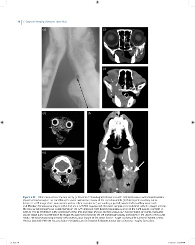
Figure 5.10 Other neoplasms of the oral cavity. (a) Osteoma. This radiograph shows a smooth proliferative mass with mineral opacity
(double‐headed arrow) on the mandible with severe periodontal disease of the rostral mandible. (b) Odontogenic maxillary tumor.
A transverse CT image shows an expansile and osteolytic mass (arrows) surrounding a partially eroded left maxillary molar tooth.
(c,d) Maxillary fibrosarcoma imaged with CT (c) and a T2W MRI sequence (d). The mass margins are less distinct in the CT images whereas
the mass with heterogeneous signal intensity in the T2W images is more distinct. Regional osteolysis of the right maxilla is present in
both, as well as infiltration in the ventral periorbital area and nasal and oral cavities (arrows). (e,f) Salivary gland carcinoma. Transverse
(e) and dorsal plane reconstruction (f) images of a carcinoma involving the left mandibular salivary gland (arrows) are shown. A metastatic
medial retropharyngeal lymph node (*) effaces the caudal margin of the tumor. Source: Images courtesy of Dr Anthony Fischetti, Animal
Medical Center, Dr Merrilee Holland, Auburn University, and Dr Shannon P. Holmes, Animal Cross‐Sectional Imaging Specialists.
c05.indd 58 1/28/2020 2:32:14 PM