
Page 67 - Feline diagnostic imaging
P. 67
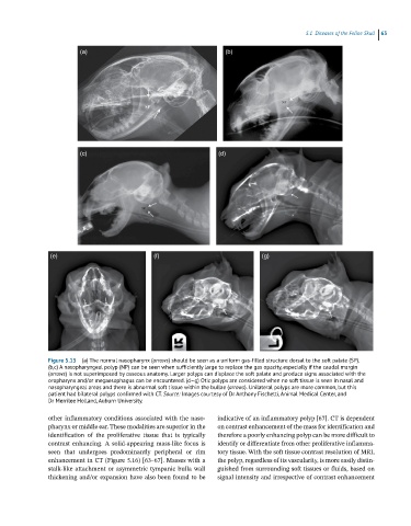
5.1  Diseases of the  eline Skull  63

                         (a)                                        (b)


                         (c)                                      (d)


                (e)                            (f)                             (g)


               Figure 5.15  (a) The normal nasopharynx (arrows) should be seen as a uniform gas-filled structure dorsal to the soft palate (SP).
               (b,c) A nasopharyngeal polyp (NP) can be seen when sufficiently large to replace the gas opacity, especially if the caudal margin
               (arrows) is not superimposed by osseous anatomy. Larger polyps can displace the soft palate and produce signs associated with the
               oropharynx and/or megaesophagus can be encountered. (d–g) Otic polyps are considered when no soft tissue is seen in nasal and
               nasopharyngeal areas and there is abnormal soft tissue within the bullae (arrows). Unilateral polyps are more common, but this
               patient had bilateral polyps confirmed with CT. Source: Images courtesy of Dr Anthony Fischetti, Animal Medical Center, and
               Dr Merrilee Holland, Auburn University.

               other inflammatory conditions associated with the naso-  indicative of an inflammatory polyp [67]. CT is dependent
               pharynx or middle ear. These modalities are superior in the   on contrast enhancement of the mass for identification and
               identification  of  the  proliferative  tissue  that  is  typically   therefore a poorly enhancing polyp can be more difficult to
               contrast  enhancing.  A  solid‐appearing  mass‐like  focus  is   identify or differentiate from other proliferative inflamma-
               seen  that  undergoes  predominantly  peripheral  or  rim   tory tissue. With the soft tissue contrast resolution of MRI,
               enhancement in CT (Figure 5.16) [63–67]. Masses with a   the polyp, regardless of its vascularity, is more easily distin-
               stalk‐like attachment or asymmetric tympanic bulla wall   guished from surrounding soft tissues or fluids, based on
               thickening and/or expansion have also been found to be   signal intensity and irrespective of contrast enhancement