
Page 68 - Feline diagnostic imaging
P. 68
64 5 Diagnostic Imaging of Diseases of the Skull
(a) (b)
(c)
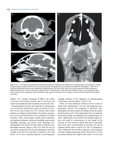
Figure 5.16 CT images in a postcontrast soft tissue window. (a) Transverse, (b) dorsal, and (c) sagittal plane reconstructions. A large
nasopharyngeal polyp (NP) fills the area, laterally expands the nasopharynx, and ventrally displaces the soft palate. A thin rim of
contrast enhancement can be seen outlining the polyp (arrows). The size of the polyp also causes narrowing of the oropharynx.
Nonenhancing fluid is seen in the medial compartment of the left bulla on the transverse image (a). This is most likely associated
with occlusion of the eustachian tube and indicates the origin of the polyp. Source: Images courtesy of Dr Merrilee Holland, Auburn
University.
(Figure 5.17). Another advantage of MRI is the ability imaging modality in the diagnosis of nasopharyngeal
to evaluate surrounding anatomy and in particular the narrowing or stenosis (Figure 5.18) [73–75].
central and peripheral nervous system structures [63, 68]. There are some important technical factors to bear in
Nasopharyngeal stenosis and imperforate nasopharynx mind when performing CT of cats with suspected naso-
is an uncommon disease that causes narrowing or occlu- pharyngeal stenosis. The diagnosis of abnormal naso-
sion of the nasal airways [69, 70]. However, since feline pharyngeal soft tissue requires air filling around the region
patients are obligate nasal breathers, the clinical presenta- for delineation on CT. The endotracheal tube can push the
tion can be severe when there is near‐complete or complete soft palate dorsally and diminish the nasopharyngeal air-
occlusion. Other clinical signs include equal inspiratory space. Additionally, mucus within the nasopharyngeal air-
and expiratory dyspnea, stertorous breathing, open‐mouth way will result in overestimation of the length of the
breathing, sneezing, and chronic nasal discharge [71]. stenosis. It is also critical to obtain thin slices (1 mm thick
Radiographs of the skull have been used to diagnose naso- slices have been recommended) to minimize the risk of
pharyngeal stenosis (Figure 5.18a) [72]. This can only be missing smaller lesion lesions [73]. The CT diagnosis is
accurately interpreted if the lateral radiograph is perfectly often confirmed with retroflex rhinoscopy and antegrade
straight, such that the X‐ray beam is parallel to the hard contrast nasopharyngoscopy, which have proven to more
palate. CT is more routinely reported as the diagnostic accurately estimate the length of the lesion. It is not only