
Page 70 - Feline diagnostic imaging
P. 70
66 5 Diagnostic Imaging of Diseases of the Skull
important to the diagnosis, but also to the more recently relation to surrounding tissues [5, 6, 65]. It has been sug-
employed interventional techniques used to treat these cats gested that CT is better for evaluating the osseous compo-
(Figure 5.18). Treatment with balloon dilation and naso- nents of this region and MRI is superior for soft tissue
pharyngeal stent placements resulted in an overall success- evaluation and delineation of abnormalities [65]. With
ful outcome in 78% of patients [76]. appropriate MR sequence selection, good detail of the
osseous structures of the ear can be achieved [79]. MRI is
also superior for assessing the endolymph within the inner
5.1.3 Ear Disease
ears, since changes in signal intensity without anatomic
Diagnostic imaging of the feline aural complex is predomi- distortion can indicate otitis interna.
nantly focused on the inner and middle ear components, Otitis or inflammatory/infectious disease processes of
since the external ear canal can be examined with otos- the feline ear have multiple causes. Scottish fold cats and
copy. Imaging of the ears is primarily undertaken for the Persians or older Siamese cats are predisposed due to
diagnosis of inflammatory or neoplastic conditions. their pinna confirmation or excessive ceruminous excre-
Historically, radiographs have been used to evaluate the tions, respectively [80]. Otitis media occurs when the
inner and middle ear regions. Low sensitivity for bulla dis- mucosal lining of tympanic cavity becomes inflamed and
ease has been reported generally, but there are also studies it is most commonly associated with respiratory disease.
that show equivalent performance of radiographs to CT in The most common underlying cause of otitis media in
the diagnoses of otitis media [4]. feline patients is polyp formation even when the polyp is
The air‐filled external ear canals are best seen and not located within the aural complex [81]. Occlusion of the
compared on a DV/VD image. The symmetry of the mid- eustachian tube is likely primarily responsible and there-
dle and inner ear regions can also be compared on these fore other disease processes involving the nasal cavities and
radiographs. However, lateral 20° ventral‐lateral dorsal nasopharynx have been associated with otitis media and/or
oblique of the left and right bulla, rostrocaudal 10° ven- abnormal fluid accumulation in the tympanic cavities
tral‐caudal dorsal oblique and a rostrocaudal open‐mouth [73, 82, 83]. Isolated otitis media (i.e., in the absence of
radiograph have been reported to better highlight the tym- otitis externa) is more common in the cat than the dog,
panic bulla (Figure 5.19) [6, 65, 77, 78]. Cross‐sectional causing soft tissue attenuation within the tympanic bulla
techniques, similar to other regions of the skull, have the or bullae (Figure 5.20) [80, 84]. Thickening of the tym-
added advantage of evaluating the entire aural complex. panic bulla is a feature of chronic otitis media, whereas
The internal otic architecture is completely visualized in osteolysis is uncommon. Intracranial otogenic extension
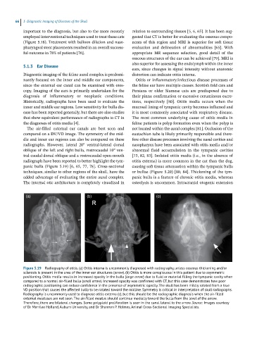
(a) (b) (c)
Figure 5.19 Radiography of otitis. (a) Otitis interna is uncommonly diagnosed with radiography, unless osseous thickening and/or
sclerosis is present in the area of the inner ear structures (arrow). (b) Otitis is more conspicuous in this patient due to asymmetric
positioning. Otitis media results in increased opacity in the bulla (large arrow) due to fluid or material filling the tympanic cavity when
compared to a normal air‐filled bulla (small arrow). Increased opacity was confirmed with CT, but this case demonstrates how poor
radiographic positioning can reduce confidence in the presence of asymmetric opacity. The skull has been mildly rotated from a true
VD position that causes the affected bulla to be rotated toward the midline. Symmetry is critical in interpretation of skull radiographs.
Radiography is uncommonly used to diagnose otitis externa (c), but this should be the radiographic diagnosis when the air‐filled
external meatuses are not seen. The air‐filled meatus should continue medially toward the bulla from the level of the arrow.
Therefore, there are bilateral changes. Some polyploid proliferation is seen in the canal lateral to the arrow. Source: Images courtesy
of Dr Merrilee Holland, Auburn University, and Dr Shannon P. Holmes, Animal Cross‐Sectional Imaging Specialists.