
Page 74 - Feline diagnostic imaging
P. 74
70 5 Diagnostic Imaging of Diseases of the Skull
(a) (b) (c)
(d) (e) (f)
(g)
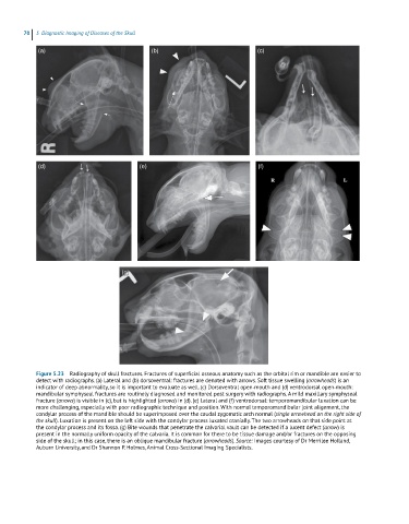
Figure 5.23 Radiography of skull fractures. Fractures of superficial osseous anatomy such as the orbital rim or mandible are easier to
detect with radiographs. (a) Lateral and (b) dorsoventral: fractures are denoted with arrows. Soft tissue swelling (arrowheads) is an
indicator of deep abnormality, so it is important to evaluate as well. (c) Dorsoventral open‐mouth and (d) ventrodorsal open‐mouth:
mandibular symphyseal fractures are routinely diagnosed and monitored post surgery with radiographs. A mild maxillary symphyseal
fracture (arrows) is visible in (c), but is highlighted (arrows) in (d). (e) Lateral and (f) ventrodorsal: temporomandibular luxation can be
more challenging, especially with poor radiographic technique and position. With normal temporomandibular joint alignment, the
condylar process of the mandible should be superimposed over the caudal zygomatic arch normal (single arrowhead on the right side of
the skull). Luxation is present on the left side with the condylar process luxated cranially. The two arrowheads on that side point at
the condylar process and its fossa. (g) Bite wounds that penetrate the calvarial vault can be detected if a lucent defect (arrow) is
present in the normally uniform opacity of the calvaria. It is common for there to be tissue damage and/or fractures on the opposing
side of the skull; in this case, there is an oblique mandibular fracture (arrowheads). Source: Images courtesy of Dr Merrilee Holland,
Auburn University, and Dr Shannon P. Holmes, Animal Cross‐Sectional Imaging Specialists.