
Page 66 - Feline diagnostic imaging
P. 66
62 5 Diagnostic Imaging of Diseases of the Skull
(a) (b) (c)
(d) (e)
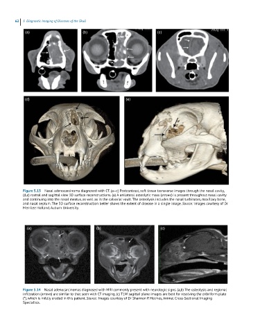
Figure 5.13 Nasal adenocarcinoma diagnosed with CT. (a–c) Postcontrast, soft tissue transverse images through the nasal cavity,
(d,e) rostral and sagittal view 3D surface reconstructions. (a) A unilateral osteolytic mass (arrows) is present throughout nasal cavity
and continuing into the nasal meatus, as well as in the calvarial vault. The osteolysis includes the nasal turbinates, maxillary bone,
and nasal septum. The 3D surface reconstruction better shows the extent of disease in a single image. Source: Images courtesy of Dr
Merrilee Holland, Auburn University.
(a) (b) (c)
Figure 5.14 Nasal adenocarcinomas diagnosed with MRI commonly present with neurologic signs. (a,b) The osteolysis and regional
infiltration (arrows) are similar to that seen with CT imaging. (c) T1W sagittal plane images are best for resolving the cribriform plate
(*), which is mildly eroded in this patient. Source: Images courtesy of Dr Shannon P. Holmes, Animal Cross‐Sectional Imaging
Specialists.