
Page 57 - Feline diagnostic imaging
P. 57
5.1 Diseases of the eline Skull 53
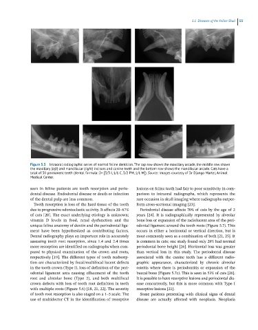
Figure 5.5 Intraoral radiographic series of normal feline dentition. The top row shows the maxillary arcade, the middle row shows
the maxillary (left) and mandibular (right) incisors and canine teeth and the bottom row shows the mandibular arcade. Cats have a
total of 30 permanent teeth (dental formula: 2× [3/3 I, 1/1 C, 3/2 PM, 1/1 M]). Source: Images courtesy of Dr Django Martel, Animal
Medical Center.
seen in feline patients are tooth resorption and perio - lesions on feline teeth had fair to poor sensitivity in com-
dontal disease. Endodontal disease or death or infection parison to intraoral radiographs, which represents the
of the dental pulp are less common. rare occasion in skull imaging where radiographs outper-
Tooth resorption is loss of the hard tissue of the tooth form cross‐sectional imaging [23].
due to progressive odontoclastic activity. It affects 20–67% Periodontal disease affects 70% of cats by the age of 2
of cats [20]. The exact underlying etiology is unknown; years [24]. It is radiographically represented by alveolar
vitamin D levels in food, renal dysfunction and the bone loss or expansion of the radiolucent area of the peri-
unique feline anatomy of dentin and the periodontal liga- odontal ligament around the tooth roots (Figure 5.7). This
ment have been hypothesized as contributing factors. occurs in either a horizontal or vertical direction, but is
Dental radiography plays an important role in accurately most commonly seen as a combination of both [21, 25]. It
assessing tooth root resorption, since 1.4 and 2.4 times is common in cats; one study found only 28% had normal
more resorption are identified on radiographs when com- periodontal bone height [26]. Horizontal loss was greater
pared to physical examination of the crown and roots, than vertical loss in this study. The periodontal disease
respectively [19]. The different types of tooth reabsorp- associated with the canine teeth has a different radio-
tion are characterized by focal/multifocal lucent defects graphic appearance, characterized by chronic alveolar
in the tooth crown (Type I), loss of definition of the peri- osteitis where there is periodontitis or expansion of the
odontal ligament area causing effacement of the tooth buccal bone (Figure 5.7c). This is seen in 53% of cats [26].
root and alveolar bone (Type 2), and both multifocal It is possible to have resorptive lesions and periodontal dis-
crown defects with loss of tooth root definition in teeth ease concurrently, but this is more common with Type I
with multiple roots (Figure 5.6) [18, 21, 22]. The severity resorptive lesions [21].
of tooth root resorption is also staged on a 1–5 scale. The Some patients presenting with clinical signs of dental
use of multidector CT in the identification of resorptive disease are actually affected with neoplasia. Neoplasia