
Page 81 - Feline diagnostic imaging
P. 81
78 6 Imaging the Feline Neurologic System
(a) (b)
(c)
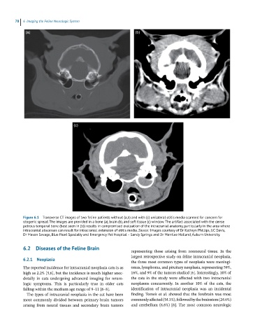
Figure 6.1 Transverse CT images of two feline patients without (a,b) and with (c) unilateral otitis media scanned for concern for
otogenic spread. The images are provided in a bone (a), brain (b), and soft tissue (c) window. The artifact associated with the dense
petrous temporal bone (best seen in (b)) results in compromised evaluation of the intracranial anatomy, particularly in the area where
intracranial abscesses can result for intracranial extension of otitis media. Source: Images courtesy of Dr Kathryn Phillips, UC Davis,
Dr Mason Savage, Blue Pearl Specialty and Emergency Pet Hospital – Sandy Springs and Dr Merrilee Holland, Auburn University.
6.2 Diseases of the Feline Brain
representing those arising from nonneural tissue. In the
largest retrospective study on feline intracranial neoplasia,
6.2.1 Neoplasia
the three most common types of neoplasia were meningi-
The reported incidence for intracranial neoplasia cats is as omas, lymphoma, and pituitary neoplasia, representing 59%,
high as 2.2% [5,6], but the incidence is much higher anec- 16%, and 9% of the tumors studied [6]. Interestingly, 10% of
dotally in cats undergoing advanced imaging for neuro- the cats in the study were affected with two intracranial
logic symptoms. This is particularly true in older cats neoplasms concurrently. In another 10% of the cats, the
falling within the medium age range of 9–11 [6–8]. identification of intracranial neoplasia was an incidental
The types of intracranial neoplasia in the cat have been finding. Tomek et al. showed that the forebrain was most
most commonly divided between primary brain tumors commonly affected (54.1%), followed by the brainstem (24.6%)
arising from neural tissues and secondary brain tumors and cerebellum (6.6%) [8]. The most common neurologic