
Page 83 - Feline diagnostic imaging
P. 83
80 6 Imaging the Feline Neurologic System
(a) (b) (c)
(d) (e) (f)
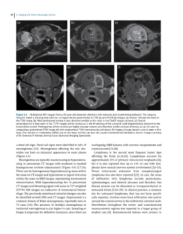
Figure 6.4 Multiplanar MR images from a 10-year-old domestic shorthair with seizures and recent hiding behavior. The imaging
features match a meningioma with iso- to hyperintense parenchyma in T2W (a) and FLAIR (b) images. (a) Arrows indicate the mass in
the T2W image. (b) Mild perilesional edema is also detected medial to the mass in the FLAIR images (arrows). (c) Central
mineralization is best seen in the T2*W images (white arrows), as is the thickening of the calvarial vault (hyperostosis) adjacent to the
lesion (black arrow). Meningiomas (white arrows) are highly vascular tumors and therefore avidly contrast enhance, as can be seen by
comparing a precontrast T1W image (d) with postcontrast T1W transverse (e) and dorsal (f) images. A large necrotic core is seen in this
mass. The midline is moderately shifted due to the mass and the cat also had caudal transtentorial herniation. Source: Images courtesy
of Dr Shannon P. Holmes, Animal Cross-Sectional Imaging Specialists.
a dural tail sign. Dural tail signs were identified in 64% of overlapping MRI features with cerebral toxoplasmosis and
meningiomas [14]. Meningiomas affecting the tela cho- cryptococcosis [19,20].
roidea can have an intraaxial appearance in some planes Lymphoma is the second most frequent tumor type
(Figure 6.5). affecting the brain [6,14,21]. Lymphomas account for
Meningiomas are typically isoattenuating to hyperattenu- approximately 35% of primary intracranial neoplasms [6],
ating in precontrast CT images with moderate to marked but it is also reported that up to 13% of cats with lym-
homogeneous contrast enhancement (Figure 6.6) [17,18]. phoma have central nervous system involvement [22–25].
There can be heterogeneous hyperattenuating areas within Direct intracranial extension from nasopharyngeal
the mass on CT images and hypointense or signal void areas lymphoma has also been reported [25]. In cats, the areas
within the mass on MRI images, representing intratumoral of infiltration with lymphoma include parenchyma,
mineralization. Mild hyperattenuating foci in precontrast leptomeninges, and choroid plexuses and therefore this
CT images and blooming signal void areas on T2*‐weighted disease process can be discussed as intraparenchymal or
(T2*W) MR images are indicative of intratumoral hemor- extraaxial forms [5,26–29]. In clinical practice, a common
rhage. The previously mentioned calvarial changes can also site for extraaxial lymphoma, that has not been specifi-
be identified on both MRI and CT images. Hyperostosis is a cally reported, involves more focal infiltration into and/or
common feature of feline meningiomas, reportedly seen in around the cranial nerves in the midventral calvarial vault.
73 cases [14]. The presence of multiple meningiomas or Distribution throughout the rostro‐ and caudotentorial
incidental meningiomas is also higher in cats. Sampling or neuroanatomic regions was reported in a larger group of
biopsy is important for definitive treatment, since there are studied cats [8]. Rostrotentorial lesions were present in