
Page 87 - Feline diagnostic imaging
P. 87
84 6 Imaging the Feline Neurologic System
(a) (c)
(b) (d)
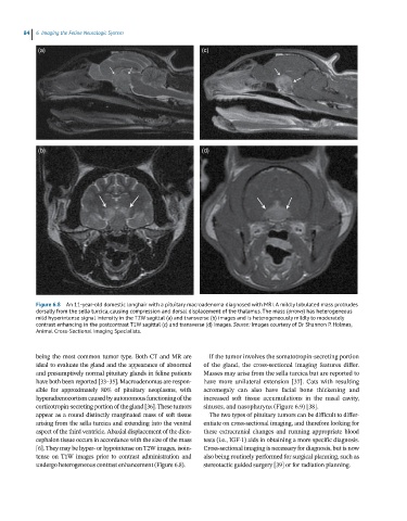
Figure 6.8 An 11-year-old domestic longhair with a pituitary macroadenoma diagnosed with MRI. A mildly lobulated mass protrudes
dorsally from the sella turcica, causing compression and dorsal displacement of the thalamus. The mass (arrows) has heterogeneous
mild hyperintense signal intensity in the T2W sagittal (a) and transverse (b) images and is heterogeneously mildly to moderately
contrast enhancing in the postcontrast T1W sagittal (c) and transverse (d) images. Source: Images courtesy of Dr Shannon P. Holmes,
Animal Cross-Sectional Imaging Specialists.
being the most common tumor type. Both CT and MR are If the tumor involves the somatotropin‐secreting portion
ideal to evaluate the gland and the appearance of abnormal of the gland, the cross‐sectional imaging features differ.
and presumptively normal pituitary glands in feline patients Masses may arise from the sella turcica but are reported to
have both been reported [33–35]. Macroadenomas are respon- have more unilateral extension [37]. Cats with resulting
sible for approximately 80% of pituitary neoplasms, with acromegaly can also have facial bone thickening and
hyperadrenocortism caused by autonomous functioning of the increased soft tissue accumulations in the nasal cavity,
corticotropin‐secreting portion of the gland [36]. These tumors sinuses, and nasopharynx (Figure 6.9) [38].
appear as a round distinctly marginated mass of soft tissue The two types of pituitary tumors can be difficult to differ-
arising from the sella turcica and extending into the ventral entiate on cross‐sectional imaging, and therefore looking for
aspect of the third ventricle. Abaxial displacement of the dien- these extracranial changes and running appropriate blood
cephalon tissue occurs in accordance with the size of the mass tests (i.e., IGF‐1) aids in obtaining a more specific diagnosis.
[6]. They may be hyper‐ or hypointense on T2W images, isoin- Cross‐sectional imaging is necessary for diagnosis, but is now
tense on T1W images prior to contrast administration and also being routinely performed for surgical planning, such as
undergo heterogeneous contrast enhancement (Figure 6.8). stereotactic guided surgery [39] or for radiation planning.