
Page 86 - Feline diagnostic imaging
P. 86
(a) (b) (c)
R R
R
(d) (e) (f)
R
R
R
(g) (h) (i)
L
R R
(j)
L
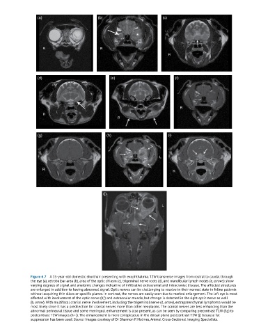
Figure 6.7 A 15-year-old domestic shorthair presenting with exophthalmia. T2W transverse images from rostral to caudal through
the eye (a), retrobulbar area (b), area of the optic chiasm (c), trigeminal nerve roots (d), and mandibular lymph nodes (e, arrows) show
varying degrees of signal and anatomic changes indicative of infiltrative extracranial and intracranial disease. The affected structures
are enlarged in addition to having abnormal signal. Optic nerves can be challenging to resolve in their normal state in feline patients
without acquiring thin slices or specific planes. In contrast, the nerves are easily seen due to marked enlargement. The left eye is most
affected with involvement of the optic nerve (b,*) and extraocular muscle, but change is detected in the right optic nerve as well
(b, arrow). With multifocal cranial nerve involvement, including the trigeminal nerve (d, arrow), extraparenchymal lymphoma would be
most likely since it has a predilection for cranial nerves more than other neoplasms. The cranial nerves are less enhancing than the
abnormal perineural tissue and some meningeal enhancement is also present, as can be seen by comparing precontrast T1W (f,g) to
postcontrast T1W images (h–j). The enhancement is more conspicuous in the dorsal plane postcontrast T1W (j) because fat
suppression has been used. Source: Images courtesy of Dr Shannon P. Holmes, Animal Cross-Sectional Imaging Specialists.