
Page 89 - Feline diagnostic imaging
P. 89
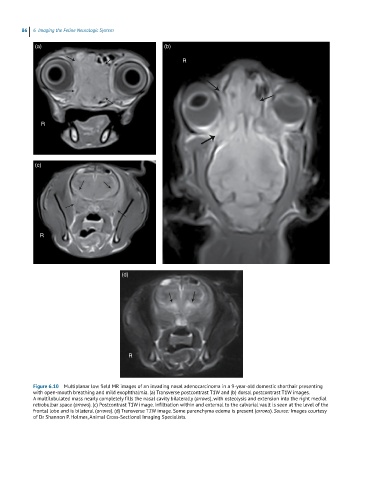
86  6  Imaging the Feline Neurologic System

            (a)                                        (b)

                                                             R


              R


            (c)


              R


                                         (d)


                                           R


            Figure 6.10  Multiplanar low field MR images of an invading nasal adenocarcinoma in a 9-year-old domestic shorthair presenting
            with open-mouth breathing and mild exophthalmia. (a) Transverse postcontrast T1W and (b) dorsal postcontrast T1W images.
            A multilobulated mass nearly completely fills the nasal cavity bilaterally (arrows), with osteolysis and extension into the right medial
            retrobulbar space (arrows). (c) Postcontrast T1W image. Infiltration within and external to the calvarial vault is seen at the level of the
            frontal lobe and is bilateral (arrows). (d) Transverse T2W image. Some parenchyma edema is present (arrows). Source: Images courtesy
            of Dr Shannon P. Holmes, Animal Cross-Sectional Imaging Specialists.