
Page 93 - Feline diagnostic imaging
P. 93
90 6 Imaging the Feline Neurologic System
(a) (b)
(c) (d)
N N
N
N
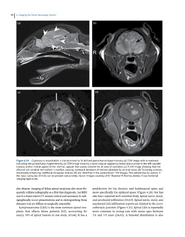
Figure 6.14 CryStococcus encephalitis is characterized by ill-defined parenchymal hyperintensity. (a) T2W image with arrowheads
indicating the parenchymal hyperintensity. (b) T2W image showing a more angular-appearing lesion (black arrow) in the left caudate
nucleus and/or medial aspect of the internal capsule that causes concern for an area of cavitation. (c) FLAIR image showing that the
affected left cerebral hemisphere is swollen, causing rightward deviation of midline (denoted by vertical lines). (d) Following contrast,
moderately enhancing multifocal intraaxial nodules (N) are identified in the postcontrast T1W images. This patient has no lesions in
the nasal cavity, but rhinitis can be present concurrently. Source: Images courtesy of Dr Shannon P. Holmes, Animal Cross-Sectional
Imaging Specialists.
disc disease. Imaging of feline spinal neoplasia also more fre- predilection for the thoracic and lumbosacral spine and
quently utilizes radiography as a first‐line diagnostic, but MRI more specifically the epidural space (Figure 6.20), but has
and to a lesser extent CT remain critical and necessary in radi- also been reported with vertebral body, spinal nerve, dural,
ographically occult presentations and in distinguishing those and arachnoid infiltration [24,63]. Spinal nerve, dural, and
diseases that are diffuse or surgically amenable. arachnoid LSA infiltration reports are limited to the cervi-
Lymphosarcoma (LSA) is the most common spinal neo- cothoracic junction (Figure 6.21). Spinal LSA is reportedly
plasia that affects feline patients [62], accounting for more common in young cats with mean ages between
nearly 39% of spinal tumors in one study [63,64]. It has a 3.6 and 5.9 years [24,63]. A bimodal distribution is also