
Page 97 - Feline diagnostic imaging
P. 97
94 6 Imaging the Feline Neurologic System
extramedullary regions. FSA has been reported to be more
common in the thoracic region and it is speculated that this
may be related to a vaccine injection site [63].
When evaluating spines with suspected neoplasia, it is
important to identify pathologic fractures which worsen
prognosis. Some may result in sufficient demineraliza-
tion and distortion to be detectable with radiography
(Figure 6.25).
6.3.2 Infection
Infectious myelitis, arachnoiditis, meningitis, and epidural
empyema are more commonly encountered than inflam-
matory conditions in feline patients. It is, however, a rela-
tively uncommon imaging diagnosis in comparison to
neoplasia, intervertebral disc disease and even vascular
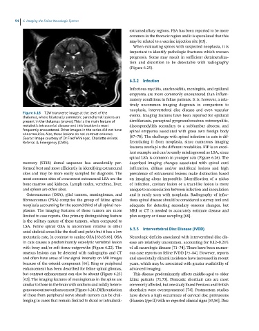
Figure 6.18 T2W transverse image at the level of the events. Imaging features have been reported for epidural
thalamus, where bilaterally symmetric parenchymal lesions are
present in the thalamus (arrows). This is the main feature of dirofilariasis, paraspinal pyogranulomatous osteomyelitis,
metabolic intracranial disease and this location is most discospondylitis secondary to a sublumbar abscess, and
frequently encountered. Other images in the series did not have spinal empyema associated with grass awn foreign body
abnormalities. Also, these lesions do not contrast enhance. [67–70]. The challenge with spinal infection in cats is dif-
Source: Image courtesy of Dr Fred Wininger, Charlotte Animal
Referral & Emergency (CARE). ferentiating it from neoplasia, since numerous imaging
features overlap in the different modalities. FIP is an excel-
lent example and can be easily misdiagnosed as LSA, since
spinal LSA is common in younger cats (Figure 6.26). The
recovery (STIR) dorsal sequence has anecdotally per- described imaging changes associated with spinal cord
formed best and most efficiently in identifying extraneural infiltration, diffuse and/or multifocal lesions and high
sites and may be more easily sampled for diagnosis. The prevalence of extraneural lesions make distinction based
most common sites of concurrent extraneural LSA are the on imaging alone impossible. Identification of a nidus
bone marrow and kidneys. Lymph nodes, vertebrae, liver, of infection, cavitary lesion or a tract‐like lesion is more
and spleen are other sites. unique to an association between infection and inoculation
Osteosarcoma (OSA), glial tumors, meningiomas, and and is rarely seen with neoplasia. Radiography of infec-
fibrosarcomas (FSA) comprise the group of feline spinal tious spinal disease should be considered a survey tool and
neoplasia accounting for the second third of all spinal neo- adequate for detecting secondary osseous changes, but
plasms. The imaging features of these tumors are more MRI or CT is needed to accurately estimate disease and
limited to case reports. One primary distinguishing feature plan surgery or tissue sampling [68].
is the solitary nature of these tumors, when compared to
LSA. Feline spinal OSA is uncommon relative to other 6.3.3 Intervertebral Disc Disease (IVDD)
axial skeletal areas like the skull and pelvis but it has a low
metastatic rate, in contrast to canine OSA [63,65,66]. OSA Neurologic deficits associated with intervertebral disc dis-
in cats causes a predominantly osteolytic vertebral lesion ease are relatively uncommon, accounting for 0.12–0.24%
with bony and/or soft tissue outgrowths (Figure 6.22). The of all neurologic disease [71–74]. There have been numer-
osseous lesions can be detected with radiographs and CT ous case reports on feline IVDD [75–84]. However, reports
and often have areas of low signal intensity on MR images and anecdotally clinical incidence have increased in recent
because of the osteoid component [66]. Ring or peripheral years, which may be associated with greater availability of
enhancement has been described for feline spinal gliomas, advanced imaging.
but contrast enhancement can also be absent (Figure 6.23) This disease predominantly affects middle‐aged to older
[63]. The imaging features of meningiomas in the spine are feline patients [71,73]. Domestic shorthair cats are most
similar to those in the brain with uniform and mildly hetero- commonly affected, but one study found Persians and British
geneous contrast enhancement (Figure 6.24). Differentiation shorthairs were overrepresented [74]. Postmortem studies
of these from peripheral nerve sheath tumors can be chal- have shown a high occurrence of cervical disc protrusions
lenging in cases that remain limited to dural or intradural‐ (Hansen type II) with no reported clinical signs [85,86]. Disc