
Page 102 - Feline diagnostic imaging
P. 102
6.3 iseases oo the Feline SSine 99
(a) (b)
(c)
(d) (e)
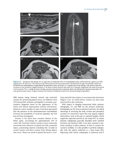
Figure 6.23 Multiplanar MR images of a 11-year-old cat diagnosed with an intramedullary mass, confirmed to be a glioma. (a) T2W
sagittal, (b) STIR dorsal, (c) T2W transverse, (d) T1W transverse, and (e) postcontrast T1W transverse images. Arrowheads denote the
ill-defined and heterogeneous hyperintense intramedullary mass at caudal L1 in sagittal and dorsal images, with arrows indicating
the mass on the transverse images. Portions of the mass contrast enhance and these are irregularly marginated. This area of the spinal
cord is swollen. The L1 vertebral body is focally narrowed and smoothly contoured, which was attributed to pressure resorption of a
slow growing mass. Source: Images courtesy of Dr Shannon P. Holmes, Animal Cross-Sectional Imaging Specialists.
MRI lesions, being bilateral, ventral, and territorial tions and soft tissue injury to neuromuscular structures
around the penetrating spinal artery, are different from (Figure 6.32). As noted earlier, trauma can also cause
FCE lesions [96]. Ischemic myelopathy is routinely a pre - intervertebral disc extrusion.
sumptive diagnosis, based on the appearance of the With respect to imaging traumatized feline patients,
lesion and clinical improvement without intervention. radiographs, CT, and MRI are the primary modalities.
Based on a small number of cases, it has been speculated Radiographs are the most economical and have the added
that the presence of the aforementioned comorbidities advantage of surveying the body cavities and paraspinal
increases the possibility of recurrent episodes, but this anatomy for concurrent injury that may require surgical
has not been investigated. intervention, such as free gas or ruptured bladder, which
Trauma is the third most common disease of the negatively impacted survival in one study [97]. In canine
feline spine, accounting for approximately 14% of patients, radiography generally identifies most vertebral
cases [94]. Vertebral column trauma is most commonly fractures and luxations [98]; it has been assumed it per-
associated with vehicular accidents but falls, projectile forms similarly in feline patients. If there is concern for
injury, penetrating injuries including bite wounds in excessive mobility, it is also possible to perform radiogra-
animal attacks and blunt trauma from falling objects phy with the patient stabilized to a back board [99].
also occur. These can result in spinal fractures or luxa- Beginning with lateral radiographs is preferred and if