
Page 100 - Feline diagnostic imaging
P. 100
(f) (g) (h)
(i) (j)
Figure 6.20 (Continued)
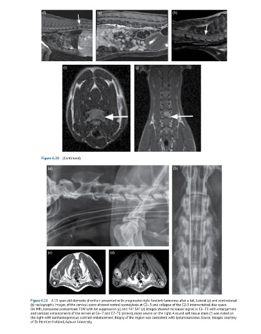
(a) (b)
(c) (d)
Figure 6.21 A 15-year-old domestic shorthair presented with progressive right forelimb lameness after a fall. Lateral (a) and ventrodorsal
(b) radiographic images of the cervical spine showed ventral spondylosis at C2–3 and collapse of the C2-3 intervertebral disc space.
On MRI, transverse postcontrast T1W with fat suppression (c) and FAT SAT (d) images showed increased signal at C6–T1 with enlargement
and contrast enhancement of the nerves at C6–7 and C7–T1 (arrows), more severe on the right. A round soft tissue mass (*) was noted on
the right with nonhomogeneous contrast enhancement. Biopsy of the region was consistent with lymphosarcoma. Source: Images courtesy
of Dr Merrilee Holland, Auburn University.