
Page 103 - Feline diagnostic imaging
P. 103
(a) (c)
(b)
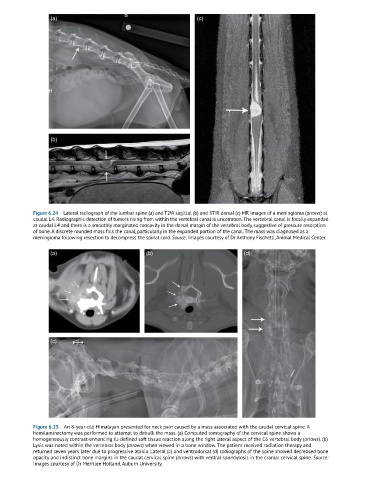
Figure 6.24 Lateral radiograph of the lumbar spine (a) and T2W sagittal (b) and STIR dorsal (c) MR images of a meningioma (arrows) at
caudal L4. Radiographic detection of tumors rising from within the vertebral canal is uncommon. The vertebral canal is focally expanded
at caudal L4 and there is a smoothly marginated concavity in the dorsal margin of the vertebral body, suggestive of pressure resorption
of bone. A discrete rounded mass fills the canal, particularly in the expanded portion of the canal. The mass was diagnosed as a
meningioma following resection to decompress the spinal cord. Source: Images courtesy of Dr Anthony Fischetti, Animal Medical Center.
(a) (b) (d)
(c)
Figure 6.25 An 8-year-old Himalayan presented for neck pain caused by a mass associated with the caudal cervical spine. A
hemilaminectomy was performed to attempt to debulk the mass. (a) Computed tomography of the cervical spine shows a
homogeneously contrast-enhancing ill-defined soft tissue reaction along the right lateral aspect of the C6 vertebral body (arrows). (b)
Lysis was noted within the vertebral body (arrows) when viewed in a bone window. The patient received radiation therapy and
returned seven years later due to progressive ataxia. Lateral (c) and ventrodorsal (d) radiographs of the spine showed decreased bone
opacity and indistinct bone margins in the caudal cervical spine (arrows) with ventral spondylosis in the cranial cervical spine. Source:
Images courtesy of Dr Merrilee Holland, Auburn University.