
Page 104 - Feline diagnostic imaging
P. 104
6.3 iseases oo the Feline SSine 101
(a) (b)
(c) (d)
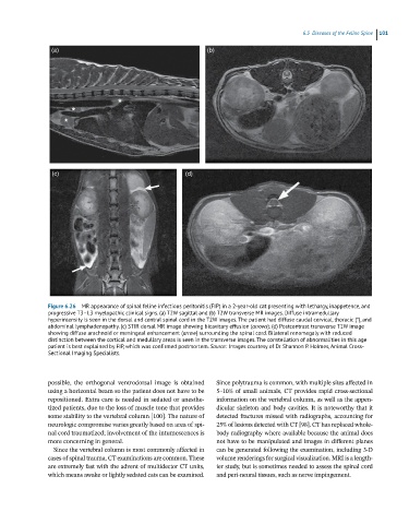
Figure 6.26 MR appearance of spinal feline infectious peritonitis (FIP) in a 2-year-old cat presenting with lethargy, inappetence, and
progressive T3–L3 myelopathic clinical signs. (a) T2W sagittal and (b) T2W transverse MR images. Diffuse intramedullary
hyperintensity is seen in the dorsal and central spinal cord in the T2W images. The patient had diffuse caudal cervical, thoracic (*), and
abdominal lymphadenopathy. (c) STIR dorsal MR image showing bicavitary effusion (arrows). (d) Postcontrast transverse T1W image
showing diffuse arachnoid or meningeal enhancement (arrow) surrounding the spinal cord. Bilateral renomegaly with reduced
distinction between the cortical and medullary areas is seen in the transverse images. The constellation of abnormalities in this age
patient is best explained by FIP, which was confirmed postmortem. Source: Images courtesy of Dr Shannon P. Holmes, Animal Cross-
Sectional Imaging Specialists.
possible, the orthogonal ventrodorsal image is obtained Since polytrauma is common, with multiple sites affected in
using a horizontal beam so the patient does not have to be 5–10% of small animals, CT provides rapid cross‐sectional
repositioned. Extra care is needed in sedated or anesthe- information on the vertebral column, as well as the appen-
tized patients, due to the loss of muscle tone that provides dicular skeleton and body cavities. It is noteworthy that it
some stability to the vertebral column [100]. The nature of detected fractures missed with radiographs, accounting for
neurologic compromise varies greatly based on area of spi- 25% of lesions detected with CT [98]. CT has replaced whole‐
nal cord traumatized; involvement of the intumescences is body radiography where available because the animal does
more concerning in general. not have to be manipulated and images in different planes
Since the vertebral column is most commonly affected in can be generated following the examination, including 3‐D
cases of spinal trauma, CT examinations are common. These volume renderings for surgical visualization. MRI is a length-
are extremely fast with the advent of multidector CT units, ier study, but is sometimes needed to assess the spinal cord
which means awake or lightly sedated cats can be examined. and peri‐neural tissues, such as nerve impingement.