
Page 109 - Feline diagnostic imaging
P. 109
106 6 Imaging the Feline Neurologic System
(a) (b)
SC
(c) (d) (e)
M M
SC
(f) (g)
M
M M M
L
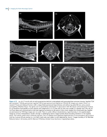
Figure 6.32 (a–d) A 7-month-old cat with progressive decline to tetraplegia with presumed but unknown trauma. Sagittal T2W
(a), transverse T2W (b), postcontrast sagittal T1W (c), and postcontrast transverse T1W (d) MR images are shown. There is a
caudal C7 body and physeal fracture with a large heterogeneously hyperintense and enhancing early fibrous callus and/or
surrounding inflamed tissue. The spinal cord is secondarily compressed in association with the tissue surrounding the fracture.
(e–g) Traumatic myelopathy without spinal cord compression in a 3-year-old cat that was caught in a garage door. The cat did
not have deep pain at presentation, but had regained sensation to the pelvic limbs when imaged the subsequent day. Sagittal
(e) and transverse T2W (f,g) images are included. There is subtle diffuse intramedullary hyperintensity in the sagittal plane
image (e, arrows). Interpretation of this change is challenging due to inappropriate phase encoding direction in the sagittal
plane. The lumbar spinal cord is diffusely swollen. There is marked heterogeneous hyperintensity in the paraspinal musculature
and the muscles (M) are swollen (e–g). A left-sided body wall defect is also detected (g). Source: Images courtesy of Dr Merrilee
Holland, Auburn University, and Dr Shannon P. Holmes, Animal Cross-Sectional Imaging Specialists.