
Page 107 - Feline diagnostic imaging
P. 107
104 6 Imaging the Feline Neurologic System
(a) (a)
(b)
(b)
(c)
(c)
(d)
(d)
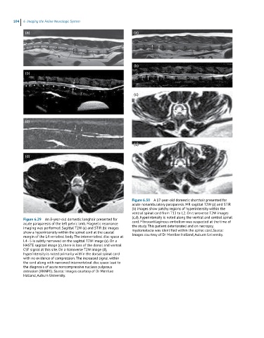
Figure 6.30 A 17-year-old domestic shorthair presented for
acute nonambulatory paraparesis. MR sagittal T2W (a) and STIR
(b) images show patchy regions of hyperintensity within the
ventral spinal cord from T13 to L2. On transverse T2W images
(c,d), hyperintensity is noted along the ventral and central spinal
Figure 6.29 An 8-year-old domestic longhair presented for cord. Fibrocartilaginous embolism was suspected at the time of
acute paraparesis of the left pelvic limb. Magnetic resonance the study. This patient deteriorated and on necropsy,
imaging was performed. Sagittal T2W (a) and STIR (b) images myelomalacia was identified within the spinal cord.Source:
show a hyperintensity within the spinal cord at the caudal Images courtesy of Dr Merrilee Holland, Auburn University.
margin of the L4 vertebral body. The intervertebral disc space at
L4–5 is subtly narrowed on the sagittal T2W image (a). On a
HASTE sagittal image (c), there is loss of the dorsal and ventral
CSF signal at this site. On a transverse T2W image (d),
hyperintensity is noted primarily within the dorsal spinal cord
with no evidence of compression. The increased signal within
the cord along with narrowed intervertebral disc space lead to
the diagnosis of acute noncompressive nucleus pulposus
extrusion (ANNPE). Source: Images courtesy of Dr Merrilee
Holland, Auburn University.