
Page 106 - Feline diagnostic imaging
P. 106
6.3 iseases oo the Feline SSine 103
(a) (b) (c)
SC SC
(d) (e)
(f) (g)
SC
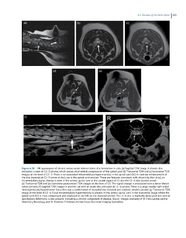
Figure 6.28 MR appearance of chronic versus acute intervertebral disc herniation in cats. (a) Sagittal T2W image. A chronic disc
extrusion is seen at C2–3 (arrow), which causes mild ventral compression of the spinal cord. (b) Transverse T2W and (c) transverse T1W
images at the level of C2–3. There is no associated intramedullary hyperintensity in the spinal cord (SC) or contrast enhancement of
the disc material at C2–3 (arrow at disc), nor is the spinal cord swollen. These are features consistent with chronicity. Also in (a), an
intramedullary signal change is seen in the ventral spinal cord at the caudal aspect of C5 and the C5–6 disc (curved arrow).
(d) Transverse T2W and (e) transverse postcontrast T1W images at the level of C5. The signal change is associated with a nerve sheath
tumor (arrows). (f) Sagittal T2W images in another cat with an acute disc extrusion at L5–6 (arrow). There is a large, mostly right-sided
heterogeneously hyperintense focus that was a combination of extruded disc material and epidural steatitis (arrow). (g) Transverse T2W
image at the level of L5–6. Focal intramedullary hyperintensity is present in the central spinal cord in the transverse image where the
spinal cord (SC) is most compressed and displaced to the left by disc material (arrow). The L5–6 disc is markedly desiccated and ventral
spondylosis deformans is also present, indicating a chronic component of disease. Source: Images courtesty of Dr Chris Levine, Levine
Veterinary Neurology, and Dr Shannon P. Holmes, Animal Cross-Sectional Imaging Specialists.