
Page 108 - Feline diagnostic imaging
P. 108
6.3 iseases oo the Feline SSine 105
(a) (b) (c)
(d) (e) (f)
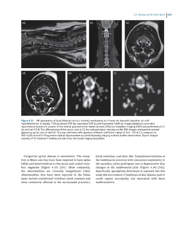
Figure 6.31 MR appearance of focal bilateral cervical ischemic myelopathy in a 9-year-old domestic shorthair cat with
hyperthyroidism. In sagittal T2W (a), dorsal STIR (b), transverse T2W (c), and transverse FLAIR (d) images, bilaterally symmetric
hyperintense lesions are present in the ventral gray and white matter (arrows). Diffusion-weighted imaging (DWI) was performed at C2
(e) and mid C4 (f). The affected area of the spinal cord at C2 has reduced signal intensity on the DWI images compared to normal-
appearing spinal cord at mid C4. This was confirmed with apparent diffusion coefficient values of 650–750 at C2, compared to
960–1120 at mid C4. Progressive clinical improvement occurred following imaging without further intervention. Source: Images
courtesy of Dr Shannon P. Holmes, Animal Cross-Sectional Imaging Specialists.
Congenital spinal disease is uncommon. The excep - block vertebrae, and extra ribs. Transitional vertebrae at
tion is Manx cats that have been reported to have spina the lumbosacral junction with concurrent asymmetry in
bifida and hemivertebrae in the sacral and caudal verte - the sacroiliac joints predispose cats to degenerative disc
bral segments (Figure 6.33) [101]. More commonly, changes at the lumbosacral joint (Figure 6.34) [102].
the abnormalities are clinically insignificant [102]. Specifically, spondylosis deformans is reported, but this
Abnormalities that have been reported in the feline study did not evaluate if lumbosacral disc disease and/or
spine include transitional vertebrae (most common and cauda equine neuropathy was associated with these
most commonly affected at the sacrocaudal junction), malformations.