
Page 105 - Feline diagnostic imaging
P. 105
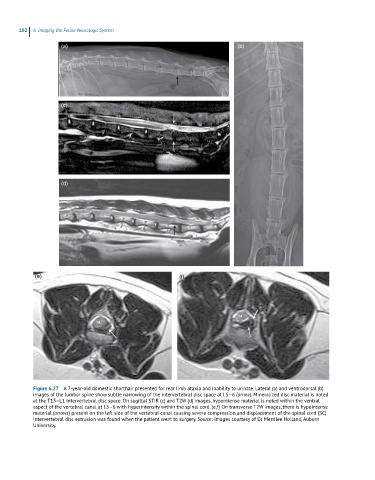
102  6  Imaging the Feline Neurologic System

                     (a)                                                       (b)


                     (c)


                     (d)


            (e)                                             (f)


            Figure 6.27  A 7-year-old domestic shorthair presented for rear limb ataxia and inability to urinate. Lateral (a) and ventrodorsal (b)
            images of the lumbar spine show subtle narrowing of the intervertebral disc space at L5–6 (arrow). Mineralized disc material is noted
            at the T13–L1 intervertebral disc space. On sagittal STIR (c) and T2W (d) images, hypointense material is noted within the ventral
            aspect of the vertebral canal at L5–6 with hyperintensity within the spinal cord. (e,f) On transverse T2W images, there is hypointense
            material (arrows) present on the left side of the vertebral canal causing severe compression and displacement of the spinal cord (SC).
            Intervertebral disc extrusion was found when the patient went to surgery. Source: Images courtesy of Dr Merrilee Holland, Auburn
            University.