
Page 110 - Feline diagnostic imaging
P. 110
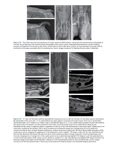
(a)                            (b)              (d)


                              (c)


               Figure 6.33  Two Manx cats with varying degrees of caudal vertebral malformations. Lateral (a) and ventrodorsal (b) radiographs of
               the first cat and lateral (c) and ventrodorsal (d) radiographs of the second cat are characterized by misshapen caudal vertebrae,
               scoliosis, and kyphosis. The second cat also had a chronic fracture of the right femur. Neither cat had associated neurologic deficits,
               but there can be issues associated with this malformation. Source: Images courtesy of Dr Merrilee Holland, Auburn University.

                            (a)                        (b)                 (c)


                            (d)


                                                                           (g)


                            (e)


                                                                           (h)
                            (f)


               Figure 6.34  A 7-year-old domestic shorthair presented for reluctance to jump onto the furniture. On the lateral (a) and ventrodorsal
               (b) radiographs of the pelvis, the L7–S1 intervertebral disc space appears narrowed and wedge shaped. There was a questionable
               ventral step defect of L7 relative to L6. There is also a vestigial disc space at S1–2. On a stress flexed projection (c), the intervertebral
               disc space widens and appears normal compared to the neutral and extended (d) views. On the ventrodorsal image (b), the
               intervertebral disc space is angled and there is asymmetry of the ilial wings. The malformation of the sacral region is likely due to the
               transitional vertebra at this location. MRI is useful in assessing lumbosacral disease and distinguishing clinically significant
               lumbosacral intervertebral disease. Dynamic lumbosacral disease can be documented with MR which allows better evaluation of the
               cauda equina nerves. Compare the appearance of the lumbosacral joint in sagittal T2W images made with the hips extended (e) and
               with the hips flexed (f). With flexion, the disc herniation at the lumbosacral joint no longer protrudes into the epidural space and
               therefore compression or impingement of the cauda equine nerves is alleviated. (g) Sagittal T2W image of a cat with spondylothesis
               or mild ventral subluxation of the cranial sacrum relative to caudal L7 (i.e., step misalignment). (h) Sagittal T2W image of another cat
               showing a large accumulation of extruded disc material (*) in the epidural space dorsal to the lumbosacral disc. Source: Images
               courtesy of Dr Merrilee Holland, Auburn University, and Dr Shannon P. Holmes, Animal Cross-Sectional Imaging Specialists.