
Page 101 - Feline diagnostic imaging
P. 101
98 6 Imaging the Feline Neurologic System
(a) (b)
(c) (d)
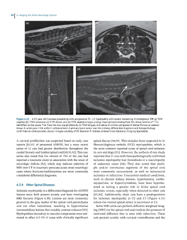
Figure 6.22 A 13-year-old Siamese presenting with progressive T2–L3 myelopathy with recent worsening. In multiplanar MR (a) T2W
sagittal, (b) T2W transverse, (c) STIR dorsal, and (d) T1W sagittal images, a large mass (arrows) arising from the dorsal lamina of T7 is
identified as the cause. The mass has low signal intensity on T2W images, indicative of a mass composed of dense fibrous or osteoid
tissue. It undergoes mild uniform enhancement. A primary bone tumor was the primary differential diagnosis and histopathology
confirmed an osteosarcoma. Source: Images courtesy of Dr Shannon P. Holmes, Animal Cross-Sectional Imaging Specialists.
A cervical predilection was suspected based on early case spinal disease [94,95]. This includes those suspected to be
reports [81,91] of presumed ANNPE, but a more recent fibrocartilaginous embolic (FCE) myelopathies, which is
series of 11 cats had greater distribution throughout the the most common reported cause of spinal cord ischemia
caudal thoracic and lumbar spinal cord [81,91,92]. This case in cats and dogs [93]. However, the authors of one study
series also noted that the owners of 75% of the cats had reported that 11 cats with histopathologically confirmed
reported a traumatic event in association with the onset of ischemic myelopathy had thrombosis or a vasculopathy
neurologic deficits [92], which may indicate selection of of unknown cause [94]. They also noted that multi-
MRI over CT in traumatic peracute/acute onset neurologic ple and/or continuous segments of the spinal cord
cases where fractures/(sub)luxations are more commonly were commonly encountered, as well as intracranial
considered differential diagnoses. ischemia or infarction. Concurrent medical conditions,
such as chronic kidney disease, hypertension, cardio -
myopathies, or hyperthyroidism, have been hypothe -
6.3.4 Other Spinal Diseases
sized as having a greater role in feline spinal cord
Ischemic myelopathy is a differential diagnosis for ANNPE ischemic events, especially when detected in older cats
lesions since both present acutely and have overlapping [93,96]. Additionally, older cats have a predisposition
MRI features (Figure 6.30). Lesions are most commonly for ischemic myelopathy at C2 and C3 (Figure 6.31)
greatest in the gray matter of the spinal cord parenchyma where the ventral spinal artery is narrowest at C2.
and are often lateralized, resulting in hyperintense Some MRI units can perform diffusion‐weighted imag-
intramedullary lesions that variably contrast enhance [93]. ing (DWI) on the spinal cord and confirm the presence of
Myelopathies secondary to vascular compromise were esti- restricted diffusion that is seen with infarction. These
mated to affect 6.5–9% of cases with clinically significant cats present acutely with cervical ventroflexion and the