
Page 96 - Feline diagnostic imaging
P. 96
6.3 iseases oo the Feline SSine 93
(a)
(b) (c)
(d)
(e)
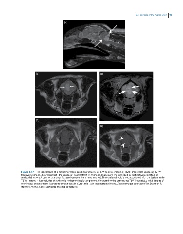
Figure 6.17 MR appearance of a nonhemorrhagic cerebellar infarct. (a) T2W sagittal image, (b) FLAIR transverse image, (c) T2*W
transverse image, (d) precontrast T1W image, (e) postcontrast T1W image. Images are characterized by distinctly marginated or
territorial lesions. A territorial margin is seen between the arrows in (a–c). Since a signal void is not associated with the lesion in the
T2*W images, it is concluded that there is no hemorrhagic component. Compared to the precontrast T1W image (d), a mild degree of
meningeal enhancement is present (arrowheads in e), but this is an inconsistent finding. Source: Images courtesy of Dr Shannon P.
Holmes, Animal Cross-Sectional Imaging Specialists.