
Page 95 - Feline diagnostic imaging
P. 95
92 6 Imaging the Feline Neurologic System
(a) (b) (c)
(d) (e) (f)
(g) (h)
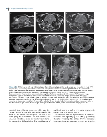
Figure 6.16 MRI images of an 8-year-old domestic shorthair with meningitis secondary to otogenic spread from otitis media. (a) T2W
sagittal image. The normally air-filled bulla is filled with tissue/material with heterogeneous signal intensity (*). (b) T2W transverse
image. There is also extension beyond the bulla into the medial aspect of the horizontal ear canal and ventral to the ear canal (arrows).
(c) FLAIR transverse image. No extension is seen from the mass (arrows) into the cranium. (d) T2*W transverse images show
heterogeneous low signal to signal void areas that are suspicious for hemorrhage (arrows). (e) Precontrast T1W transverse image.
Compare the appearance of the mass (arrows) in the precontrast image with postcontrast images. (f) Postcontrast T1W (*). (g,h)
Postcontrast T1W dorsal images. Following contrast administration, the tissue and material within and associated with the bulla undergo
moderate to no enhancement, respectively. Extensive meningeal enhancement and mild thickening are present, indicating intracranial
extension of disease as seen on (f) and (h) (arrows). The extent of disease is visible in the transverse image (*) but is best appreciated on
the dorsal plane images (arrows). Source: Images courtesy of Dr Shannon P. Holmes, Animal Cross-Sectional Imaging Specialists.
reported, thus affecting young and older cats (11– additional lesions, as well as extraneural structures, is
16 years of age). Interestingly, in reviewing cases for this another important imaging feature.
book, all the known cases of spinal LSA were in the Because of the relatively high occurrence of concurrent
older group. Multifocal lesions are more common with extraneural LSA, reportedly up to 85–100% [63], screening
LSA than other feline spinal neoplasms, which may aid MR series or widening of the CT field of view to include the
in antemortem differentiation. The identification of extraneural tissue are necessary. A short tau inversion