
Page 94 - Feline diagnostic imaging
P. 94
6.3 iseases oo the Feline SSine 91
(a) (b) (c)
(e)
(d) (f)
(g) (h)
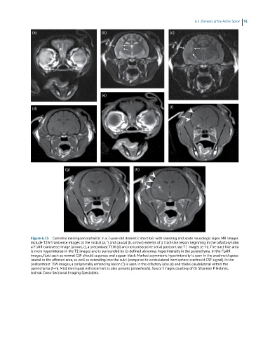
Figure 6.15 Cuterebra meningoencephalitis in a 2-year-old domestic shorthair with sneezing and acute neurologic signs. MR images
include T2W transverse images at the rostral (a, *) and caudal (b, arrows) extents of a tract-like lesion beginning in the olfactory lobe,
a FLAIR transverse image (arrows, c), a precontrast T1W (d) and nonconsecutive serial postcontrast T1 images (e–h). The tract-like area
is more hyperintense in the T2 images and is surrounded by ill-defined abnormal hyperintensity in the parenchyma. In the FLAIR
images, fluid such as normal CSF should suppress and appear black. Marked asymmetric hyperintensity is seen in the arachnoid space
lateral to the affected area, as well as extending into the sulci (compared to contralateral hemisphere arachnoid CSF signal). In the
postcontrast T1W images, a peripherally enhancing lesion (*) is seen in the olfactory lobe (e) and tracks caudolateral within the
parenchyma (f–h). Mild meningeal enhancement is also present (arrowheads). Source: Images courtesy of Dr Shannon P. Holmes,
Animal Cross-Sectional Imaging Specialists.