
Page 91 - Feline diagnostic imaging
P. 91
88 6 Imaging the Feline Neurologic System
(a) (b) (c)
(d) (f)
(e)
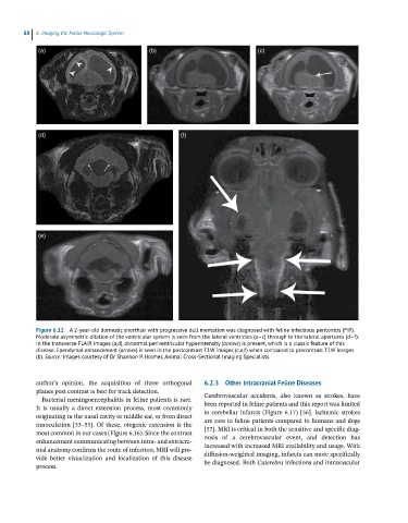
Figure 6.12 A 2-year-old domestic shorthair with progressive dull mentation was diagnosed with feline infectious peritonitis (FIP).
Moderate asymmetric dilation of the ventricular system is seen from the lateral ventricles (a–c) through to the lateral apertures (d–f).
In the transverse FLAIR images (a,d), abnormal periventricular hyperintensity (arrows) is present, which is a classic feature of this
disease. Ependymal enhancement (arrows) is seen in the postcontrast T1W images (c,e,f) when compared to precontrast T1W images
(b). Source: Images courtesy of Dr Shannon P. Holmes, Animal Cross-Sectional Imaging Specialists.
author’s opinion, the acquisition of three orthogonal 6.2.3 Other Intracranial Feline Diseases
planes post contrast is best for track detection.
Bacterial meningoencephalitis in feline patients is rare. Cerebrovascular accidents, also known as strokes, have
It is usually a direct extension process, most commonly been reported in feline patients and this report was limited
originating in the nasal cavity or middle ear, or from direct to cerebellar infarcts (Figure 6.17) [56]. Ischemic strokes
are rare in feline patients compared to humans and dogs
innoculation [53–55]. Of these, otogenic extension is the
most common in our cases (Figure 6.16). Since the contrast [57]. MRI is critical in both the sensitive and specific diag-
nosis of a cerebrovascular event, and detection has
enhancement communicating between intra‐ and extracra-
nial anatomy confirms the route of infection, MRI will pro- increased with increased MRI availability and usage. With
diffusion‐weighted imaging, infarcts can more specifically
vide better visualization and localization of this disease
process. be diagnosed. Both Cuterebra infections and intravascular