
Page 99 - Feline diagnostic imaging
P. 99
96 6 Imaging the Feline Neurologic System
(a) (b) (c)
(d) (e)
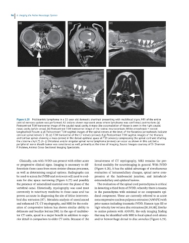
Figure 6.20 Multicentric lymphoma in a 15-year-old domestic shorthair presenting with multifocal signs. MRI of the entire
central nervous system was performed. All lesions shown represent areas where lymphoma was confirmed postmortem. (a)
Postcontrast T1W transverse image of the caudal nasal cavity. A mass-like accumulation of tissue is seen in the right caudal
nasal cavity (white arrow). (b) Postcontrast T1W transverse image of the rostral neurocranium. White arrowheads = rostral
longitudinal fissure. (c,d) Postcontrast T1W sagittal images of the spinal nerves at the level of the foramina (arrowheads indicate
cervical spinal nerves 5–8). (e) T2W transverse of the C7 nerves (arrows). (f,g) Postcontrast T1W sagittal images of the thoracic
and lumbar spines showing a mass (arrow) in the dorsal epidural space at T10 severely compressing the spinal cord and dilating
the cisterna chyli (*). (h–j) Unilateral and/or singular spinal nerve lymphoma (arrows) can occur as shown in this cat, but a
peripheral nerve sheath tumor was considered as well, primarily at the time of imaging. Source: Images courtesy of Dr Shannon
P. Holmes, Animal Cross-Sectional Imaging Specialists.
Clinically, cats with IVDD can present with either acute invasiveness of CT myelography, MRI remains the pre-
or progressive clinical signs. Imaging is necessary to dif- ferred modality for neuroimaging in general. With IVDD
ferentiate these cases from more sinister disease processes, (Figure 6.28), it has the added advantage of simultaneous
as well as determining surgical options. Radiographs can evaluation of intramedullary changes, spinal nerve com-
be used to screen for IVDD and reviewers will need to eval- pression at the lumbosacral junction, and intradural‐
uate for disc space narrowing (Figure 6.27) and possibly extramedullary and epidural lesions.
the presence of mineralized material over the plane of the The evaluation of the spinal cord parenchyma is critical
vertebral canal. Historically, myelography was used most in detecting a third form of IVDD, whereby there is trauma
commonly in veterinary medicine in these cases and has to the parenchyma with minimal or no compressive epi-
proven accurate in diagnosing the site of feline interverte- dural component. These are currently referred to as acute
bral disc extrusion [87]. Metadata analysis of unenhanced noncompressive nucleus pulposus extrusion (ANNPE) with
and enhanced CT, CT myelography, and MRI for the evalu- prior names including traumatic IVDD, Hansen type III or
ation of compressive lesions has shown similar ability to high velocity‐low volume disc extrusions [81,89,90]. Similar
delineate and localize lesions [88]. In the age of multidec- to canine patients with ANNPE, the only imaging finding
tor CT units, speed is a major benefit in addition to supe- that may be identified with MRI is focal spinal cord edema
rior detail in comparison to older CT units. Because of the and/or hemorrhage dorsal to disc annulus (Figure 6.29).