
Page 85 - Feline diagnostic imaging
P. 85
82 6 Imaging the Feline Neurologic System
(a) (b)
(c)
(d)
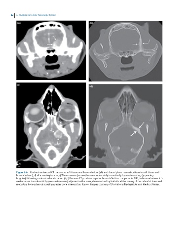
Figure 6.6 Contrast-enhanced CT transverse soft tissue and bone window (a,b) and dorsal plane reconstructions in soft tissue and
bone window (c,d) of a meningioma. (a,c) These masses (arrows) become moderately to markedly hyperattenuating (appearing
brighter) following contrast administration. (b,d) Because CT provides superior bone definition compared to MRI, in bone windows it is
easier to see the calvarial hyperostosis (arrows) adjacent to the mass, characterized by both focal thickening of the calvarial bone and
medullary bone sclerosis causing greater bone attenuation. Source: Images courtesy of Dr Anthony Fischetti, Animal Medical Center.