
Page 552 - Veterinary Immunology, 10th Edition
P. 552
initiated by multiple damage signals, including injection of
VetBooks.ir mitochondria, the formation of an apoptosome, and activation of
granzymes, and leads to the release of cytochrome C from
caspase-9.
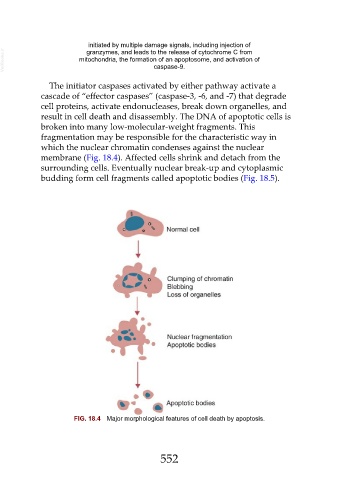
The initiator caspases activated by either pathway activate a
cascade of “effector caspases” (caspase-3, -6, and -7) that degrade
cell proteins, activate endonucleases, break down organelles, and
result in cell death and disassembly. The DNA of apoptotic cells is
broken into many low-molecular-weight fragments. This
fragmentation may be responsible for the characteristic way in
which the nuclear chromatin condenses against the nuclear
membrane (Fig. 18.4). Affected cells shrink and detach from the
surrounding cells. Eventually nuclear break-up and cytoplasmic
budding form cell fragments called apoptotic bodies (Fig. 18.5).
FIG. 18.4 Major morphological features of cell death by apoptosis.
552