
Page 553 - Veterinary Immunology, 10th Edition
P. 553
VetBooks.ir
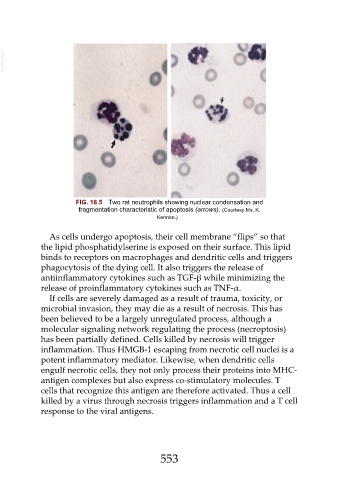
FIG. 18.5 Two rat neutrophils showing nuclear condensation and
fragmentation characteristic of apoptosis (arrows). (Courtesy Ms. K.
Kennon.)
As cells undergo apoptosis, their cell membrane “flips” so that
the lipid phosphatidylserine is exposed on their surface. This lipid
binds to receptors on macrophages and dendritic cells and triggers
phagocytosis of the dying cell. It also triggers the release of
antiinflammatory cytokines such as TGF-β while minimizing the
release of proinflammatory cytokines such as TNF-α.
If cells are severely damaged as a result of trauma, toxicity, or
microbial invasion, they may die as a result of necrosis. This has
been believed to be a largely unregulated process, although a
molecular signaling network regulating the process (necroptosis)
has been partially defined. Cells killed by necrosis will trigger
inflammation. Thus HMGB-1 escaping from necrotic cell nuclei is a
potent inflammatory mediator. Likewise, when dendritic cells
engulf necrotic cells, they not only process their proteins into MHC-
antigen complexes but also express co-stimulatory molecules. T
cells that recognize this antigen are therefore activated. Thus a cell
killed by a virus through necrosis triggers inflammation and a T cell
response to the viral antigens.
553