
Page 102 - Veterinary Histology of Domestic Mammals and Birds, 5th Edition
P. 102
84 Veterinary Histology of Domestic Mammals and Birds
VetBooks.ir
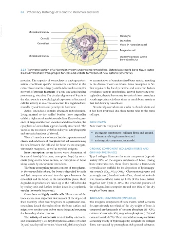
3.33 Transverse section of a Haversian system undergoing remodelling. Osteoclasts resorb bone tissue; osteo-
blasts differentiate from progenitor cells and initiate formation of new systems (schematic).
proteins. The capacity of osteoclasts to undergo polari- to accumulation of unmineralised bone matrix, resulting
sation, coordinate specific movements and bind to the in the disease known as rickets. Bone resorption is fur-
extracellular matrix is largely attributable to this complex ther regulated by local paracrine and autocrine factors
network of protein filaments (F-actin) and actin-binding (cytokines, various interleukins, growth factors and pros-
proteins (e.g. vinculin). The circular alignment of F-actin in taglandins, thyroid hormone). Per unit of time, osteoclasts
the clear zone is a morphological expression of increased resorb approximately three times as much bone matrix as
cellular activity in an active osteoclast. It is regulated hor- that laid down by osteoblasts.
monally, by calcitonin and parathyroid hormone. Structurally, osteoclasts are similar to chondroclasts and
Active osteoclasts contain abundant mitochondria. it has been proposed that these terms refer to the same
Lying internal to the ruffled border, these organelles cell type.
exhibit a high rate of aerobic metabolism. Due to the pres-
ence of large numbers of vacuoles and dense bodies, the Bone matrix
cytoplasm of osteoclasts appears loosely structured. The Bone matrix is composed of:
vacuoles are associated with the endocytic, autophagocytic
and exocytic functions of the cell. · an organic component (collagen fibres and ground
The cell membrane of osteoclasts incorporates several substance rich in glucosamine) and
classes and subclasses of receptors that aid in maintaining · an inorganic component (minerals).
the seal between the cell and the bone matrix (integrin,
vitronectin receptors), as well as myeloid antigens. ORGANIC COMPONENT (COLLAGEN FIBRES AND
Bone resorption occurs in two ways: formation of GROUND SUBSTANCE)
lacunae (Howship’s lacunae, resorption bays) by osteo- Type I collagen fibres are the main component (approxi-
clasts lying on the bone surface, or resorption of bone mately 90%) of the organic substance of bone. During
along vessels by one or more osteoclasts. bone mineralisation, these fibres provide a framework
The bone resorption process consists of two phases. (crystallisation scaffold) for the deposition of hydroxyapa-
In the extracellular phase, the bone is degraded by acids tite crystals (Ca [PO ] [OH] ). Glycosaminoglycans and
10 4 6 2
and lytic enzymes released into the space between the proteoglycans (chondroitin-4-sulfate, chondroitin-6-sul-
osteoclast and the bone. In the intracellular phase, these fate, keratin sulfate) make up 1–2% of the bone matrix.
degradation products are taken up at the ruffled border Together with lipids (5–10%), the structural proteins of
by endocytosis and further broken down in cytoplasmic the collagen fibres comprise around one third of the dry
vesicles (primarily lysosomes). weight of bone tissue.
Osteoclasts are highly mobile cells. The texture of the
bone matrix is an important determinant of the degree of INORGANIC COMPONENT (MINERALS)
their mobility. After resorbing bone in a particular area, The inorganic component of bone matrix, which accounts
osteoclasts detach themselves from the bone surface and for approximately two-thirds of the dry weight of bone, is
migrate to another area before reattaching and resuming composed predominantly of calcium phosphate (85–90%),
the bone degradation process. calcium carbonate (8–10%), magnesium phosphate (1.5%) and
The activity of osteoclasts is inhibited by calcitonin, calcium fluoride (0.3%). These minerals form a crystal lattice
and stimulated by 1,25-dihydroxycholecalciferol (vitamin (consisting mostly of hydroxyapatite) alongside the collagen
D ) and parathyroid hormone. Vitamin D deficiency leads fibres, surrounded by proteoglycan rich ground substance.
3 3
Vet Histology.indb 84 16/07/2019 14:56