
Page 105 - Veterinary Histology of Domestic Mammals and Birds, 5th Edition
P. 105
Connective and supportive tissues (textus connectivus) 87
matrix. The collagen fibres are arranged in spirals along the tion as part of lifelong bone remodelling. Loss of bone
VetBooks.ir long axis of the central canal. In alternate lamellae, the spiral mass with advancing age is associated with a reduction in
collagen fibres are oriented in different directions, forming a osteocytes and delayed mineralisation.
criss-crossing network. Cross-linkages are formed between
At the internal and external surfaces of the bone, the
adjacent lamellae. These features impart stability to the lamellae are arranged in circular sheets, forming the inter-
bone under forces of pressure and tension. nal circumferential lamellae (adjacent to the endosteum)
In additional to its structural role, lamellar bone – like and external circumferential lamellae (lying against the
all tissues derived from connective tissue – also plays a sig- internal surface of the periosteum). Collagen fibres that
nificant part in metabolism. This is enabled in large part bind the periosteum to the bone (fibrae perforantes,
by functional interactions between the bone cells, blood Sharpey’s fibres) are incorporated into the external cir-
vessels and connective tissue within the osteon. cumferential lamellae. The endosteum, composed of
Osteocytes within lacunae are regularly arranged osteoprogenitor cells and connective tissue, lines the inter-
between the concentric lamellae around the central canal nal circumferential lamellae and the trabeculae (Figure
(Figures 3.34 to 3.37). Their long cytoplasmic processes 3.34).
radiate into interconnecting canaliculi (canaliculi ossei) Trabecular (spongy) bone also contains lamellae, but
extending from the lacunae. The cellular extensions estab- these are not organised into osteonal systems. Remodelling
lish contact with the processes of other osteocytes via processes occur at a particularly high rate in spongy
gap junctions. This arrangement permits the transport bone.
of substances between the blood vessel in the Haversian
canal and the bone matrix (both to and from the vessel). New bone formation (osteogenesis)
Channels running transversely through the bone (perfo- The formation of new bone occurs in two ways.
rating canals, Volkmann’s canals) connect central canals Development of bone directly from mesenchymal con-
with each other, and with the endosteum and periosteum. nective tissue without a cartilaginous precursor phase is
Through this interconnecting network of blood vessels, referred to as intramembranous ossification (primary or
bone is a well-vascularised tissue (Figure 3.34). direct ossification). This type of bone formation gives rise
Lamellar bone serves as a metabolically active calcium to certain flat bones of the skull and the bony collar of
depot. Under the influence of parathyroid hormone, acti- developing long bones. It is also observed in repairing bone
vation of osteolytic osteocytes or, in larger areas of bone fractures. Formation of bone based on a cartilaginous tem-
resorption, multinuclear osteoclasts, within bone lacunae plate is termed endochondral ossification (secondary
2+
and canaliculi results in rapid liberation of Ca and PO or indirect ossification). This results first in (immature)
3−
4
ions from the mineralised matrix. These ions are swiftly woven bone that is gradually replaced by (mature) lamellar
conveyed to the circulation by the intraosseous transport bone (sometimes referred to as replacement bone) (Figures
system. 3.40 to 3.42).
Any change in the mechanical load on a bone results
in dynamic adaptation of its internal structure. Non- INTRAMEMBRANOUS OSSIFICATION
functional osteons are broken down, leaving remnants In intramembranous ossification, osteoprogenitor cells
referred to as interstitial lamellae (Figures 3.34 and 3.35). differentiate into osteoblasts (Figures 3.38 and 3.39) that
The structure of compact bone undergoes gradual altera- produce collagen fibres and osteoid. Through continued
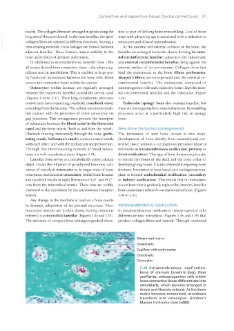
3.38 Intramembranous ossification:
bone of cranium (juvenile dog). Near
capillaries, osteoprogenitor cells within
loose connective tissue differentiate into
osteoblasts, which become arranged in
sheets and liberate osteoid. As the bone
matrix becomes mineralised, osteoblasts
transform into osteocytes. Goldner’s
Masson trichrome stain (x480).
Vet Histology.indb 87 16/07/2019 14:56