
Page 130 - Veterinary Histology of Domestic Mammals and Birds, 5th Edition
P. 130
112 Veterinary Histology of Domestic Mammals and Birds
VetBooks.ir Loose connective tissue
with collagen fibrils
Perineurium
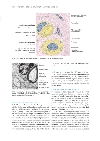
Myelinated nerve fibres
Schwann cell with nucleus
Fibrocyte within
endoneurium
Nerve fibre (transverse section)
Myelin sheath
Mesaxon
Endoneurium
Unmyelinated nerve fibres Mesaxon
Neurolemma
Schwann cell with nucleus Neurolemma
Nerve fibre (transverse section) Nerve fibre
Mesaxon (transverse section)
5.15 Structure of a myelinated and unmyelinated nerve fibre (schematic).
This layer is referred to as the sheath of Schwann (Figure
5.14).
myelination of centRal neRve fibRes
Myelination of central nerve fibres differs primarily from
that of peripheral nerve fibres in that one oligodendrocyte
ensheathes several axons (Figure 5.11). Numerous trape-
zoid processes extend from the oligodendrocyte, their free
edges becoming associated with, and wrapping around,
multiple axons. These wrappings together constitute the
myelin sheath.
COMPOSITION OF THE MYELIN SHEATH
5.16 Fine structure of a myelinated and an unmyeli- The structure of the myelin sheath resembles that of two
nated nerve fibre (transverse section) with adjacent fused unit membranes. Electron micrographs reveal the
loose connective tissue (x6000). presence of parallel, alternating lipid and protein lay-
ers. Lipids, mainly glycerine phosphatides, cholesterol
and sphingolipids, make up around 70% of myelin. The
myelination of PeRiPheRal neRve fibRes protein component (30%) includes proteolipids, glyco-
Each Schwann cell is associated with only one axon. proteins and myelin basic protein. Due to their high fat
Initially, the Schwann cell lies adjacent to the axon, sub- content, myelin sheaths can be demonstrated using lipid
sequently folding around it, displacing the axon to the staining techniques (Figure 5.20).
centre. Opposing surfaces of the plasmalemma approach Each Schwann cell or oligodendrocyte surrounds the
one another, eventually fusing to form a mesaxon (Figure axon over a distance of approximately 1 mm (Figures 5.8
5.14). The mesaxon then extends around the axon to and 5.11). Between two consecutive glial cells, the axon is
form lamellae. Initially the outer layers of plasmalemma exposed. This gap, 0.5 μm in width, is referred to as the
become fused. As the mesaxon continues to extend, and node of Ranvier (Figures 5.8 and 5.11). The section of
the lamellae are formed, the inner layers also merge. The the myelin sheath between two consecutive nodes is the
lamellae, in their entirety, constitute the myelin sheath. internode. Pockets of cytoplasm remain in the myelin
The outer portion of the Schwann cell, containing sheath where fusion of the plasma membrane of the glial
cytoplasm and the nucleus, adjoins the myelin sheath. cell is incomplete. These conical compartments, known
Vet Histology.indb 112 16/07/2019 14:57