
Page 184 - Veterinary Histology of Domestic Mammals and Birds, 5th Edition
P. 184
166 Veterinary Histology of Domestic Mammals and Birds
VetBooks.ir
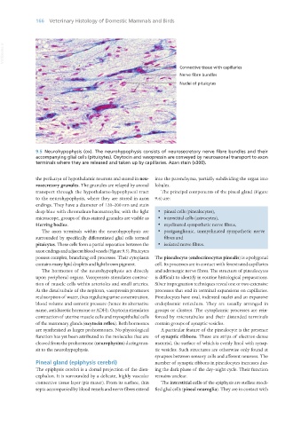
9.5 Neurohypophysis (ox). The neurohypophysis consists of neurosecretory nerve fibre bundles and their
accompanying glial cells (pituicytes). Oxytocin and vasopressin are conveyed by neuroaxonal transport to axon
terminals where they are released and taken up by capillaries. Azan stain (x300).
the perikarya of hypothalamic neurons and stored in neu- into the parenchyma, partially subdividing the organ into
rosecretory granules. The granules are relayed by axonal lobules.
transport through the hypothalamo-hypophyseal tract The principal components of the pineal gland (Figure
to the neurohypophysis, where they are stored in axon 9.6) are:
endings. They have a diameter of 120–200 nm and stain
deep blue with chromalaun-haematoxylin; with the light · pineal cells (pinealocytes),
microscope, groups of thus-stained granules are visible as · interstitial cells (astrocytes),
Herring bodies. · myelinated sympathetic nerve fibres,
The axon terminals within the neurohypophysis are · postganglionic, unmyelinated sympathetic nerve
surrounded by specifically differentiated glial cells termed fibres and
pituicytes. These cells form a partial separation between the · isolated nerve fibres.
axon endings and adjacent blood vessels (Figure 9.5). Pituicytes
possess complex, branching cell processes. Their cytoplasm The pinealocyte (endocrinocytus pinealis) is a polygonal
contains many lipid droplets and light brown pigment. cell. Its processes are in contact with fenestrated capillaries
The hormones of the neurohypophysis act directly and adrenergic nerve fibres. The structure of pinealocytes
upon peripheral organs. Vasopressin stimulates contrac- is difficult to identify in routine histological preparations.
tion of muscle cells within arterioles and small arteries. Silver impregnation techniques reveal one or two extensive
At the distal tubule of the nephron, vasopressin promotes processes that end in terminal expansions on capillaries.
reabsorption of water, thus regulating urine concentration, Pinealocytes have oval, indented nuclei and an expansive
blood volume and osmotic pressure (hence its alternative endoplasmic reticulum. They are usually arranged in
name, antidiuretic hormone or ADH). Oxytocin stimulates groups or clusters. The cytoplasmic processes are rein-
contraction of uterine muscle cells and myoepithelial cells forced by microtubules and their distended terminals
of the mammary glands (oxytocin reflex). Both hormones contain groups of synaptic vesicles.
are synthesised as larger prohormones. No physiological A particular feature of the pinealocyte is the presence
function has yet been attributed to the molecules that are of synaptic ribbons. These are strips of electron-dense
cleaved from the prohormone (neurophysins) during tran- material, the surface of which is evenly lined with synap-
sit to the neurohypophysis. tic vesicles. Such structures are otherwise only found at
synapses between sensory cells and afferent neurons. The
Pineal gland (epiphysis cerebri) number of synaptic ribbons in pinealocytes increases dur-
The epiphysis cerebri is a dorsal projection of the dien- ing the dark phase of the day–night cycle. Their function
cephalon. It is surrounded by a delicate, highly vascular remains unclear.
connective tissue layer (pia mater). From its surface, thin The interstitial cells of the epiphysis are stellate modi-
septa accompanied by blood vessels and nerve fibres extend fied glial cells (pineal neuroglia). They are in contact with
Vet Histology.indb 166 16/07/2019 14:59