
Page 188 - Veterinary Histology of Domestic Mammals and Birds, 5th Edition
P. 188
170 Veterinary Histology of Domestic Mammals and Birds
dium-like processes. A well-developed rough endoplasmic sis. These hormones are released into the basal cytoplasm
VetBooks.ir reticulum and abundant free ribosomes render the cyto- from which they pass into the capillaries.
plasm weakly basophilic. The epithelial cells are connected
by junctional complexes, separating the follicle lumen C cells (cellula parafollicularis)
from the interstitium. At their basolateral surface, epithe- Parafollicular cells, or C cells, are found individually or in
lial cells take up amino acids, monosaccharides and iodide groups in the epithelium of thyroid follicles (Figure 9.9).
(via an iodine pump) from the surrounding capillaries. These are located inside the basal lamina of the follicle
Thyroglobulin is synthesised in the rough endoplasmic but do not extend to the follicle lumen. C cells are argyro-
reticulum (Figure 9.10), then glycosylated and packaged philic and can be identified using immunohistochemical
into secretory vesicles in the Golgi apparatus. The contents techniques. Their cytoplasm stains only weakly in rou-
of the vesicles are released by exocytosis into the follicular tine preparations and thus appears pale; with electron
lumen, where they are stored. microscopy, numerous endocrine granules are visible. The
Within the colloid, iodide is oxidised to iodine (cata- granules contain the peptide hormone calcitonin and, in
lysed by membrane-bound thyroperoxidase). Iodine is smaller quantities, somatostatin, dopamine and serotonin.
complexed with free tyrosine group residues of thy- The release of calcitonin is controlled by plasma calcium
roglobulin. Coupling of iodotyrosyl residues results in the concentrations.
formation of tri-iodothyronine and tetraiodothyronine.
These are stored as inactive hormones within the thyroid Species variation
follicle. In birds, reptiles, amphibians and fish, C cells form a
The follicular epithelial cells are activated by thy- separate organ, the ultimobranchial body, that origi-
roid-stimulating hormone (TSH), produced by the nates from the epithelium of the fifth pharyngeal pouch.
adenohypophysis, and by autonomic nerves. Hormone
synthesis, storage and release (Figure 9.10) are all under Parathyroid gland (glandula
the influence of TSH. In response to a decrease in the parathyroidea)
plasma concentration of thyroxin, TSH stimulates the The small parathyroid glands lie near the thyroid gland.
resorption of the thyroglobulin–hormone complex from Septa comprising a lattice of fibres extend from the thin
the follicle into the follicular cells. This process is initi- connective tissue capsule into the interior of the gland; in
ated by liquefaction of the colloid at the perimeter of the the ox and pig this results in distinct lobulation. In carni-
follicle. The colloid is then taken up by endocytosis into vores, the septa are so insubstantial that the gland consists
phagosomes which fuse with lysosomes to form phago- essentially of parenchymal cells and capillaries. The capil-
lysosomes. Within the phagolysosomes, tri-iodothyronine laries have an irregularly expanded lumen and a fenestrated
and thyroxin are cleaved from thyroglobulin by hydroly- endothelium (sinusoids).
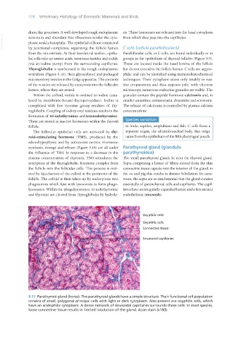
9.11 Parathyroid gland (horse). The parathyroid glands have a simple structure. Their functional cell population
consists of small, polygonal principal cells with light or dark cytoplasm. Also present are oxyphilic cells, which
have an acidophilic cytoplasm. A dense network of sinusoidal capillaries surrounds these cells. In most species,
loose connective tissue results in limited lobulation of the gland. Azan stain (x150).
Vet Histology.indb 170 16/07/2019 14:59