
Page 189 - Veterinary Histology of Domestic Mammals and Birds, 5th Edition
P. 189
Endocrine system (systema endocrinum) 171
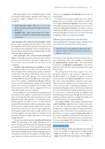
The parenchyma of the parathyroid gland consists thus acts as an antagonist of calcitonin produced by the
VetBooks.ir of relatively small polygonal epithelial cells (Figure 9.11) thyroid gland.
Parathyroid hormone acts rapidly upon bone, stimu-
arranged in cords or clusters. Two types of cells are
lating osteocytes to mobilise calcium from the surface of
recognised:
their lacunae (osteocytic osteolysis). It also exerts a long-
· small principal (chief) cells that occur in two acting effect by promoting the formation and activity of
functional stages exhibiting either light or dark cyto- osteoclasts (osteoclastic osteolysis). In addition, parathy-
plasm, and roid hormone inhibits calcium excretion in the kidney and
· oxyphilic cells – larger than principal cells, charac- stimulates absorption of calcium by the intestine through
terised by acidophilic cytoplasm with a fine granular its regulatory effect on vitamin D metabolism.
substructure.
Adrenal gland (glandula suprarenalis)
Light principal cells (endocrinocyti principales lucidi) The adrenal gland is a vital, paired organ consisting of two
have a round, centrally positioned nucleus. They contain embryologically and functionally distinct regions:
few organelles and secretory granules (resting form) but
store large amounts of glycogen. Most of the glycogen par- · adrenal cortex (cortex glandulae suprarenalis) and
ticles are dissolved during routine formol fixing, and thus · adrenal medulla (medulla glandulae suprarenalis)
these cells appear distinctly pale. (Figures 9.12 to 9.14).
Dark principal cells (endocrinocyti principales
densi) contain large quantities of electron-dense secretory The adrenal cortex develops from the mesodermal coe-
granules (secretory form). Abundant rough endoplas- lomic epithelium, while the medulla is derived from
mic reticulum and mitochondria are also present in the the neuroectoderm (neural crest). The medulla thus
cytoplasm. constitutes a sympathetic paraganglion composed of
Oxyphilic cells (endocrinocyti oxyphilici) are larger neurosecretory cells that release transmitters into capil-
than principal cells (22 μm in the ox, 27 μm in the horse). lary networks.
The chromatin of the centrally placed nucleus is often dis- The arrangement of the blood supply to the adre-
tinctly dense. The cells are filled almost exclusively with nal gland is related to the functional organisation of
mitochondria and a little glycogen. The mitochondria the gland (Figure 9.14). Beneath the capsule, an arterial
are responsible for the granular light microscopic appear- plexus branches into cortical sinusoids that extend into
ance of the cytoplasm. A transitional cell type exhibiting the cortex along with delicate connective tissue septa.
features of both principal and oxyphil cells may also be These fenestrated capillaries are accompanied by a nar-
present. The function of oxyphilic cells is unclear; it has row perivascular space containing macrophages of the
been proposed that these may be a degenerative form of mononuclear phagocyte system. At the junction between
cell as their numbers increase with age. the cortex and medulla, the capillaries open into a network
Species differences are observed in the arrangement of of venules located within the medulla. In addition, small
parenchymal cells, capillaries and perivascular connective non-branching arterioles pass through the cortex into the
tissue. In the dog, the cells of the parenchyma lie in cords medulla (medullary arterioles) to supply the capillaries of
along the usually wide capillaries. In transverse section, the medulla directly with oxygenated blood.
this arrangement resembles a rosette. In other domestic The venous vessels of the medulla converge to form
mammals, the parenchymal cells are predominantly organ- central medullary veins that leave the gland as the adre-
ised in groups or clusters and the capillaries are narrower. nal veins. The adventitia of the central medullary veins
In carnivores and in the ox, the histological appearance incorporates a longitudinal cushion of muscle which has a
of the gland is dark as the cells are small, and the nuclei regulatory effect on blood flow. The medulla thus receives
are close together. A greater volume of cytoplasm in the blood from two sources: an arterial supply, independent of
principal cells imparts a lighter appearance to the tissue in the cortical vasculature, and blood from the cortical sinu-
the pig and horse. soids, which is high in cortical hormones (corticosteroids).
The parathyroid gland produces parathyroid hormone Corticosteroids regulate the synthesis of adrenaline in the
(PTH). The ribosomes of the rough endoplasmic reticu- chromaffin cells of the medulla.
lum of the principal cells produce the large pre-hormone
pre-parathyroid hormone. This molecule is shortened by Species variation
proteases during its passage through the endoplasmic retic- Lower vertebrates: In fish and amphibians, the adrenal
ulum, before being glycosylated in the Golgi apparatus and cortex and medulla are separated into an inter-renal
released by exocytosis into the capillaries. Parathyroid hor- organ (homologous to the cortex) and an adrenal
mone increases calcium concentration in the blood and organ (chromaffin body, homologous to the medulla).
Vet Histology.indb 171 16/07/2019 14:59