
Page 360 - Veterinary Histology of Domestic Mammals and Birds, 5th Edition
P. 360
342 Veterinary Histology of Domestic Mammals and Birds
The epidermis makes up the cornified hoof capsule. As absence of secondary laminae in the wall segment.
VetBooks.ir determined by the surface architecture of the dermis, the (Figure 15.30). The epidermal hoof capsule is composed
horn of the perioplic and coronary segments, sole, frog of a dorsal border, abaxial and axial walls, the sole and
and bulbs is tubular in form. The tubules run parallel to digital pad (bulb segment). In accordance with the mecha-
the surface of the hoof. Comprising horn of suprapapil- nical forces experienced by the hoof, the tubular horn is
lar and peripapillar origin, these are firmly connected by most prominent at the perioplic and coronary segments.
intertubular horn arising between adjacent papillae. The Laminar horn is present at the bearing edge, where the
tubules extend distally from the coronary segment over wall segment transitions to the sole.
the lamellar horn of the walls and bars of the hoof. The The structure of the tubular horn differs markedly
perioplic horn forms a thin, shiny layer on the external from the layered arrangement of equines. Instead, the
surface of the hoof. tubules are arranged around the medulla in concave
The cortex of the tubules comprises concentric layers in segments, resembling the structure of a pine cone. The
which inner, middle and outer zones can be distinguished. bundles of tonofibrils are arranged in a circular fashion.
The individual layers are reinforced by tonofibrils arranged
in oppositely oriented spirals. Claw (unguicula)
Corresponding to its dermal template, the cornified The epidermal modifications that constitute the claw,
epidermis of the walls and bars is laminar in form. The or nail, are relatively simple. The perioplic and coronary
epidermis that interdigitates with the secondary dermal segments (Figure 15.31), located within the sulcus ungui-
laminae is not cornified (see Veterinary Anatomy of Domestic cularis, are underlain by a thin subcutis. The wall and sole
Mammals: Textbook and Colour Atlas). segments and their associated corium directly overlie the
unguicular process of the distal phalanx. The perioplic seg-
Ruminant and swine hoof (ungula) ment, located adjacent to the internal surface of the bony
The structure of the hoofs of ruminants and pigs exhibits ungual crest, gives rise to the external layer of claw horn.
species-specific modifications of the periople, coronary This lacks tubular horn and is relatively soft, becoming
segment, walls, sole and bulbs. Cushions formed by accu- worn away well before reaching the distal tip of the nail.
mulation of connective tissue in the subcutis protrude to The horn that develops over the papilla-studded dermis
a varying degree in the perioplic and coronary segment of the coronary segment is tubular and makes up the bulk
and in the bulbs. Particularly in the bulbs, the cushions are of the wall of the nail (claw plate) (Figure 15.31). In the
reinforced with aggregates of adipose tissue. In the wall wall segment, the corium carries very short laminae that
and sole segments, the dermis lies directly adjacent to the interdigitate with uncornified epidermal laminae. Thus,
phalanx, without a subcutis. the horn of the wall does not have a laminar structure.
The corium bears tufts of numerous short papillae. In The narrow sole segment lines the plantar surface of the
the perioplic segment these measure 1–2 mm in length. unguicular process. The epidermis of the sole is soft and
A characteristic feature of the hoof of ruminants is the non-tubular.
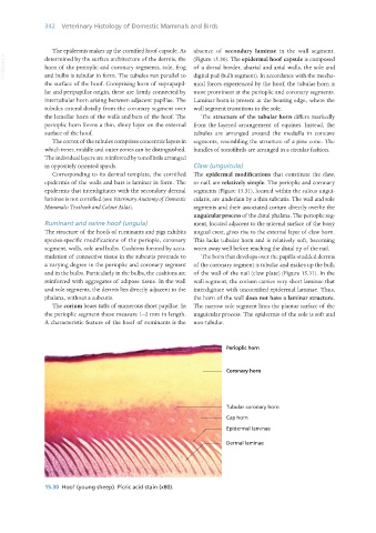
15.30 Hoof (young sheep). Picric acid stain (x80).
Vet Histology.indb 342 16/07/2019 15:06