
Page 356 - Veterinary Histology of Domestic Mammals and Birds, 5th Edition
P. 356
338 Veterinary Histology of Domestic Mammals and Birds
VetBooks.ir
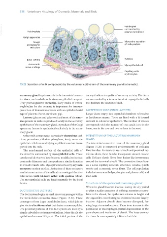
15.22 Secretion of milk components by the columnar epithelium of the mammary gland (schematic).
mammary gland by plasma cells in the interstitial connec- duct epithelium is capable of secretory activity. The ducts
tive tissue, and reach the milk via trans-epithelial transport. are surrounded by a loose network of myoepithelial cells
They provide passive immunity. Early intake of immu- that facilitate the ejection of milk.
noglobulins by the neonate is important for immune
protection of domestic mammals with an epitheliochorial LACTIFEROUS SINUS (SINUS LACTIFER)
type of placenta (horse, ruminant, pig). Larger ducts empty into expanded chambers referred to
Lactose (glucose and galactose) and most of the enzy- as lactiferous sinuses. These are lined with a bi-layered
mes present in milk are produced locally in the secretory cuboidal to columnar epithelium. The number of sinuses
epithelium of the mammary gland. A product of the Golgi corresponds with the number of teat canals (two in the
apparatus, lactose is synthesised exclusively in the mam- horse, one in the cow and two to three in the sow).
mary gland.
Other milk components, particularly electrolytes (cal- INTERSTITIUM OF THE LACTATING MAMMARY
cium, potassium, chloride, phosphate, iron), enter the GLAND
epithelial cells from underlying capilllaries and are trans- The interstitial connective tissue of the mammary gland
ported into the milk. (Figure 15.20) is composed predominantly of collagen
The non-luminal surface of the epithelial cells of fibre bundles. Particularly near alveoli and proximal lac-
the alveoli is surrounded by myoepithelial cells. These tiferous ducts, these bundles incorporate smooth muscle
ectodermal derivatives have become modified to include cells. Delicate elastic fibres form basket-like investments
contractile filaments and thus perform a similar function around the terminal alveoli. The connective tissue hou-
to smooth muscle cells. Myoepithelial cells carry oxytocin ses a dense capillary network, arterioles, venules, lymph
receptors on their surface. Activation of these receptors vessels and autonomic nerve fibres. The cell population
results in contraction of the cell and narrowing of the alve- includes immune cells (lymphocytes and plasma cells) and
lolar lumen (milk let-down reflex, milk ejection reflex). mast cells.
The myoepithelial cells are lined externally by the basal
lamina. Structure of the quiescent mammary gland
When the gland becomes inactive, during the dry period
DUCTS (DUCTUS LACTIFERI) or after a sudden cessation of milking, secretion accumu-
The duct system begins as small excretory passages within lates in the alveoli, the epithelium reduces in height and
the intralobular connective tissue (Figure 15.20). These the organelles contributing to secretion become largely
converge to form larger interlobular ducts, which join to inactive. Adjacent alveoli often become disrupted, for-
give rise to a lactiferous duct that drains a mammary lobe. ming larger terminal sections. There is an increase in the
The proximal portions of this duct system are lined by a population of macrophages, partial degeneration of the
simple cuboidal to columnar epithelium. More distally the parenchyma and involution of alveoli. The loose connec-
epithelium becomes bi-layered. The initial portion of the tive tissue becomes partially infiltrated with fat.
Vet Histology.indb 338 16/07/2019 15:06