
Page 353 - Veterinary Histology of Domestic Mammals and Birds, 5th Edition
P. 353
Common integument (integumentum commune) 335
VetBooks.ir
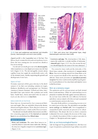
15.16 Hair and epidermal and dermal root sheaths 15.17 Skin and sinus hair (sinusoidal type, dog).
(horse). Haematoxylin and eosin stain (x250). Haematoxylin and eosin stain (x30).
aligned parallel to the longitudinal axis of the hair. This Carnivores and pigs: The distal portion of the sinus,
fibrous sheath contains blood vessels and myelinated nerve nearest the epidermis, is annular and free of trabeculae
fibres, the latter passing into the external root sheath as (sinusoid type) (Figure 15.17). Here the internal dermal
free nerve endings. root sheath bulges into the lumen of the sinus (sinus pad).
The dermal root sheath gives rise to the dermal papilla,
which manifests as a cone-shaped protrusion into the hair The connective tissue walls and the trabeculae of the
bulb (Figure 15.14). The dermal papilla contains dense blood sinus contain a fine meshwork of sensory nerve
capillary loops that supply the metabolically active cells fibres. Free nerve endings extend from these fibres as far
of the stratum basale, thereby supporting the growth and as the external root sheath of the sinus hairs, where they
development of new hairs. arborise. These nerve processes are specialised pressure
receptors that detect and transmit amplified signals gener-
Types of hair ated by perturbation of the sinus hairs and impulse waves
Domestic mammals exhibit great diversity in the devel- in the blood sinus.
opment of the hair coat including variation in length,
thickness, distribution and arrangement (see Veterinary Skin as a sensory organ
Anatomy of Domestic Mammals: Textbook and Colour Atlas). The epidermis and the nervous system are both derived
Specialised hair types include cilia, eyelashes and tactile from ectodermal tissue. While the epidermal layer serves
hairs. Tactile hairs, closely associated with the sense of primarily to insulate the body against external influences,
touch, are also referred to as sinus hairs. the nervous tissue within the skin infiltrates this barrier
and allows various stimuli to be detected and transmitted
SINUS HAIR (PILUS TACTILIS) to central sensory centres in the brain.
Sinus hairs are characterised by their pronounced thick- Receptive fields and their corresponding neurons occur
ness and length. They are embedded deep in the dermis, in various layers of the skin. The epithelium contains free
almost reaching the subcutis. There, they are in direct con- nerve endings and Merkel cells. Non-encapsulated or
tact with striated muscle, which enables their movement. encapsulated tactile corpuscles are present in the subepi-
The dermal root sheath exhibits characteristic modifica- thelial layers. The deeper the level at which a sensation
tion comprising separation of the internal and external is detected, the more complex the structure of the recep-
layers by an endothelium-lined blood sinus. tor (a detailed description of skin receptors is provided in
Chapter 16, ‘Receptors and sense organs’).
Species variation
Horse and ruminants: The entire blood sinus is tra- Skin as an immunological barrier
versed by delicate connective tissue trabeculae, dividing Like the lungs and the gastrointestinal tract, the skin is an
the sinus into irregular compartments (cavernous type) organ that presents a particularly large surface area and is
(Figure 15.18).
Vet Histology.indb 335 16/07/2019 15:06