
Page 352 - Veterinary Histology of Domestic Mammals and Birds, 5th Edition
P. 352
334 Veterinary Histology of Domestic Mammals and Birds
composed of the shaft (scapus pili) and its free tip (apex The internal root sheath is composed of three layers.
VetBooks.ir pili). The cuticle of the internal root sheath consists of a single
The medulla (medulla pili) arises from the deep lay-
layer of flattened cells that lies adjacent to the surface of
ers of the stratum basale overlying the tip of the dermal the hair. These keratinised cells are arranged in a shingle-
papilla (suprapapillar) (Figure 15.14). These epithelial cells like pattern with their free margins directed towards the
are constantly dividing, are vacuolated and are minimally bulb. The interlocking of these cells with those of the hair
keratinised. Production of new medullary cells pushes cuticle holds the hair in place.
older cells towards the exterior. The latter lose their pig- The cuticle of the root sheath is surrounded by one
mentation, their nuclei become pyknotic and trichohyalin to three layers of nucleated cells (Huxley layer) that may
granules appear within them. Degenerated medullary cells contain trichohyalin in places. A usually single layer of
may contain gas bubbles that, together with lack of pig- non-nucleated cells (Henle layer) forms the outermost
mentation, result in greying of the hair. The medulla is layer of the internal root sheath (Figure 15.16).
absent in wool fibres. The external epithelial root sheath represents the con-
The cortex (cortex pili) develops from epithelial cells tinuation of the layers of the epidermal stratum basale,
located at the margins of the papilla (peripapillar). It forms which it largely resembles in structure.
a densely cellular, strongly keratinised mantle around On the outside of the external root sheath is a clearly
the medulla. Some of its cells contain melanin pigment developed basal lamina (glassy membrane). This
granules. The density of melanosomes determines the separates the epidermal portions of the hair from the sur-
colour of the hair. Cortical keratinocytes are flattened and rounding connective tissue (mesodermal) layers.
are reinforced by longitudinal tonofilaments that impart Free sensory nerve endings penetrate the basal lamina
mechanical strength, flexibility and elasticity to the hairs. and arborise in the outer root sheath, particularly near the
The external surface of the hair is lined by a single layer follicular orifice. These nerve endings terminate in recep-
of flattened keratinised cells termed the cuticle (cuticula tors that detect touch, pressure and vibration, forming part
pili). The cells partially overlap in a manner resembling of the superficial sensory apparatus.
roof tiles, with their free edges directed towards the
hair tip (Figure 15.19). Interlocking of these cells with DERMAL ROOT SHEATH
oppositely oriented superficial cells of the internal epi- During embryonic development, the downgrowth of epi-
thelial root sheath (see below) serves to secure the hair dermal tissue into the underlying connective tissue gives
in situ. rise to a mantle termed the dermal root sheath (Figures
The structure of the medulla and cuticle is highly spe- 15.14 to 15.16). This is typically composed of two layers:
cies-specific and can thus be used for forensic purposes. an internal layer comprising collagen and elastic fibre
Distinguishing characteristics include the structure of the bundles arranged in a predominantly circular orientation,
medullary cells, the thickness and number of the medul- and an external layer in which the fibre bundles are mostly
lary layers, the relative thickness of the medulla and cortex
and the surface profile of the cells of the cuticle.
Root sheath structure
The root sheaths surrounding the hair are comprised of
the:
· epithelial root sheath:
− internal epithelial root sheath,
− external epithelial root sheath,
− basal lamina (glassy membrane),
· dermal root sheath:
− internal dermal root sheath,
− external dermal root sheath and
• dermal papilla.
EPITHELIAL ROOT SHEATH
Proximally, the epithelial root sheath forms a sleeve around
the hair, accompanying it as far as the follicular orifice at
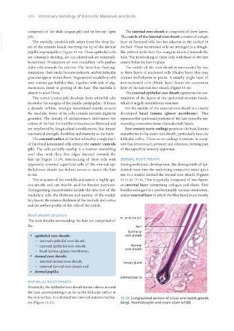
the skin surface. It is divided into internal and external lay- 15.15 Longitudinal section of a hair and sweat glands
ers (Figure 15.14). (dog). Haematoxylin and eosin stain (x100).
Vet Histology.indb 334 16/07/2019 15:06