
Page 354 - Veterinary Histology of Domestic Mammals and Birds, 5th Edition
P. 354
336 Veterinary Histology of Domestic Mammals and Birds
insult. Its lipid components, produced by the sebaceous
VetBooks.ir glands (see above), prevent the ingress of water and associ-
ated derangement of intradermal ion balance. Free fatty
acids formed during decomposition of surface lipid exert
antimicrobial effects. Both of these features serve as non-
specific protective barriers.
The epithelium incorporates Langerhans cells.
These cells are components of the mononuclear phago-
cytic system. They develop from macrophages and
migrate into the epidermis. Langerhans cells are anti-
gen-presenting cells. Antigens that have penetrated
the layers of the epidermis (e.g. bacteria) are processed
within the cell and displayed on the cell surface.
Within local lymph nodes, these antigens are presented
by Langerhans cells to T lymphocytes, which then undergo
activation and proliferation as part of the adaptive immune
response (see Chapter 8, ‘Immune system and lymphatic
organs’). The cascade of events involved in the immune
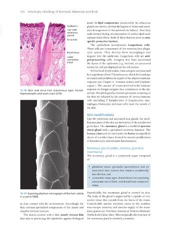
15.18 Skin and sinus hair (cavernous type, horse). response to foreign antigens thus commences in the epi-
Haematoxylin and eosin stain (x16). dermis. Morphologically, immune processes occurring in
the skin are reflected by the presence of various immune
cells (including T lymphocytes, B lymphocytes, mac-
rophages, histiocytes and mast cells) near the vessels of
the skin.
Skin modifications
Like the epidermis and associated skin glands, the modi-
fied structures of the skin are derivatives of the ectodermal
germ layer. The mammary gland is a modified apocrine
sweat gland with a specialised secretory function. The
hooves, claws and (in ruminants) the horns are superficial
sheets of cornified tissue formed by massive proliferation
of keratinocytes and extensive keratinisation.
Mammary gland (udder, mamma, glandula
mammaria)
The mammary gland is a compound organ composed
of:
· glandular tissue (glandular parenchyma) and an
associated duct system that empties peripherally
into the teat, and
· connective tissue septa (interstitium) incorporating
autonomic nerve fibres, vessels and loose connective
tissue.
15.19 Scanning electron micrograph of the hair cuticle Superficially, the mammary gland is covered in skin.
in a cat (x1500). The body of the gland is supported by a capsule of con-
nective tissue that extends from the fascia of the trunk.
in close contact with the environment. Accordingly, the Considerable species variation exists in the number,
skin contains specialised components of the innate and macroscopic anatomy and vascular supply of the mam-
adaptive immune systems. mary glands (see Veterinary Anatomy of Domestic Mammals:
The skin is covered with a thin, poorly viscous film Textbook and Colour Atlas). Microscopically, the structure of
that aids in protecting the epidermis against biological the mammary gland is relatively consistent.
Vet Histology.indb 336 16/07/2019 15:06