
Page 400 - Veterinary Histology of Domestic Mammals and Birds, 5th Edition
P. 400
382 Veterinary Histology of Domestic Mammals and Birds
interspersed between cell bodies consists of cellular dorsalis) is present at the level of the dorsal nerve roots
VetBooks.ir processes of neurons and glial cells and is referred to as (radices dorsales) of the spinal nerves. A considerably
neuropil.
shallower depression is evident at the ventral nerve roots
The marginal layer constitutes the fibre-rich white (radices ventrales).
matter, consisting predominantly of myelinated nerve
processes. Neuroglia of the white matter include oligo- Grey matter (substantia grisea)
dendrocytes, fibrous astrocytes and microglia. The lighter In transverse section, the grey matter exhibits an ‘H-shaped’
colour of the white matter is attributable to the myelin profile formed by relatively narrow dorsal horns (cornu
sheaths (formed by oligodendrocytes) surrounding the dorsale) and typically larger ventral horns (cornu ventrale)
nerve processes. (Figures 17.3 and 17.4). The horns are connected by the
lateral intermediate substance (pars intermedia lateralis).
Spinal cord (medulla spinalis) In the thoracolumbar region, the lateral intermediate sub-
The characteristic ‘H-shape’ of the grey matter (substan- stance protrudes into the white matter as the lateral horn
tia grisea) is appreciable macroscopically in cross-section (cornu lateralis). In the cervical spinal cord, an additional
of the spinal cord. Passing through the centre of the grey cellular network, the spinal reticular formation (formatio
matter is the central canal (canalis centralis), the remnant reticularis), is present between the dorsal and ventral horns.
of the lumen of the embryonic neural tube. The white The two symmetrical halves of the grey matter of the
matter (substantia alba) surrounds the grey matter, giving spinal cord are connected by a thin bridge (commissura gri-
the spinal cord its external shape (Figures 17.2 and 17.3). sea). The commissure encloses the central canal (canalis
The spinal cord is permeated by a dense capillary centralis) (Figure 17.5), which is lined by ependymal cells
network, accompanied by loose connective tissue. This (see Chapter 5, ‘Nervous tissue’). Surrounding the central
microvasculature results in fine segmentation of the nerv- canal is a ring of tissue, the central intermediate sub-
ous tissue. Highly vascular connective tissue sheaths, the stance, composed of neurons and glial cells.
meninges, surround the spinal cord. When regarded in three dimensions, the dorsal, ventral
The segments of the spinal cord (cervical, thoracic, and lateral horns form columns of tissue – the dorsal col-
lumbar, sacral) exhibit species-specific variation in the ratio umn (cornu dorsale), lateral column (cornu laterale) and
of grey to white matter and in cross-sectional area (and ventral column (cornu ventrale).
thus in volume). The grey matter is composed predominantly of
Despite these regional differences, the basic structure multipolar neurons and glial cells (astrocytes). Neuron
of the spinal cord is relatively consistent (Figure 17.2). size, number and morphology varies with location in the
The spinal cord is a bilaterally symmetrical organ, different segments of the spinal cord. Unmyelinated axons
divided by a deep ventral median fissure (fissura mediana and dendrites (neurofibrae nonmyelinata) and astrocyte
ventralis) and a shallow dorsal sulcus (sulcus medianus processes form a dense nervous tissue meshwork (neu-
dorsalis). The latter continues into the tissue of the spinal ropil). The overarching purpose of the neurons of the
cord as the dorsal median septum (septum medianum grey substance is to establish a synaptic interconnection
dorsale). A bilateral dorsolateral sulcus (sulcus lateralis between afferent and efferent neuronal pathways.
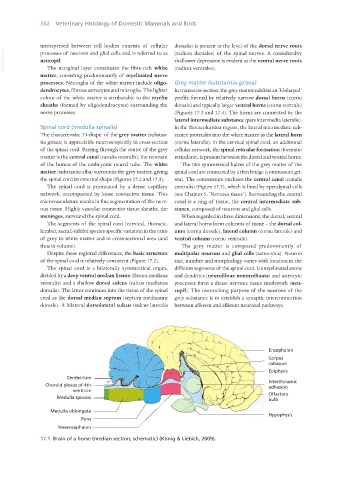
Encephalon
Corpus
callosum
Epiphysis
Cerebellum
Choroid plexus of 4th Interthalamic
adhesion
ventricle
Medulla spinalis Olfactory
bulb
Medulla oblongata
Hypophysis
Pons
Mesencephalon
17.1 Brain of a horse (median section; schematic) (König & Liebich, 2009).
Vet Histology.indb 382 16/07/2019 15:08