
Page 396 - Veterinary Histology of Domestic Mammals and Birds, 5th Edition
P. 396
378 Veterinary Histology of Domestic Mammals and Birds
VetBooks.ir
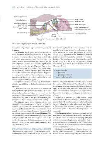
16.43 Spiral organ (organ of Corti; schematic).
three structurally different regions: vestibular, outer and duct (ductus cochlearis). Its inner section (nearest the
tympanic. modiolus) rests against a raised layer of connective tissue
The vestibular region (paries vestibularis ductus coch- (spiral limbus) on the lamina spiralis ossea. A relatively
learis, vestibular [Reissner’s] membrane) is very thin. stiff, gelatinous glycoprotein-rich membrane (tectorial
It consists of a narrow fibrous layer lined on both sides membrane, membrana tectoria) extends from the epithe-
with simple squamous epithelium. The membrane is in lial edge of the spiral limbus over the surface of the spiral
contact with the perilymph of the scala vestibuli and the organ (Figures 16.42 and 16.43). The space thus formed
endolymph of the cochlear duct. The outer region (paries is the internal spiral sulcus. The spiral organ contains the
externus) is formed by the spiral ligament (ligamentum following cell types:
spirale). Constituting an extension of the basilar mem-
brane (see below), the spiral ligament spreads out over the · supporting cells,
bone of the cochlea and is firmly bound with the perios- − pillar cells,
teum (Figure 16.40). Parts of the spiral ligament are richly − phalangeal cells and
vascularised. In the outer region, the cochlear duct is lined · sensory cells (hair cells).
by irregular pseudostratified epithelium.
The epithelial cells are characterised by abundant mito- Inner and outer pillar cells are separated by a space termed
chondria and infoldings of the basal plasmalemma (basal the inner tunnel (Corti’s tunnel). The pillar cells are flanked
striations). by phalangeal cells (inner phalangeal cells lie on the inner
A particular feature of this region is the presence of aspect of the inner pillar cells; outer phalangeal cells lie
intra-epithelial capillaries (stria vascularis). These con- on the outer side of the outer pillar cells) (Figure 16.43).
tribute to the synthesis and release of endolymph into the Between the outer pillar and phalangeal cells is the space
+
+
cochlear duct. Maintenance of low Na /high K ion con- of Nuel.
centrations is important for perception of auditory signals. The pillar cells (modified epithelial cells) have a broad
Disruption of the ion balance interferes with hearing. base that rests on the basilar membrane. Their apical cyto-
The tympanic region (paries tympanicus) is the most plasm is filled with tonofibrils (supporting fibrils). Towards
extensively differentiated portion of the wall of the coch- the epithelial surface, the pillar cells become more slender.
lear duct. The connective tissue foundation of the tympanic A characteristic plate is formed at the apices of the cells.
region is the basilar membrane (lamina basilaris), which The terminal plates of the inner and outer pillar cells are
extends from the lamina spiralis ossea to the outer wall of connected.
the cochlea, where it merges with the spiral ligament. The The phalangeal cells are arranged in a single row of
collagen fibres of the basilar membrane gradually increase inner cells and two to five rows of outer cells. These extend
in length and breadth from the base to the apex of the towards the lumen to embrace the sensory cells (hair
cochlea. Resting upon the basilar membrane is a layer of cells). Processes extending from the apices of the phalan-
modified epithelium, the spiral organ (organ of Corti) geal cells form plates that join with the hair cells and plates
(Figures 16.42 and 16.43). of the pillar cells to form the mosaic-like reticular lamina
(membrana reticularis).
SPIRAL ORGAN (ORGANUM SPIRALE, ORGAN OF CORTI) Adjoining the outer phalangeal cells are columnar outer
The spiral organ (Figure 16.43) follows a winding course, limiting cells (Hensen’s cells). Further outward, the cells
extending throughout most of the length of the cochlear become cuboidal (external supporting cells, Claudius cells)
Vet Histology.indb 378 16/07/2019 15:08