
Page 393 - Veterinary Histology of Domestic Mammals and Birds, 5th Edition
P. 393
Receptors and sense organs (organa sensuum) 375
Internal ear (auris interna) SACCULUS AND UTRICULUS
VetBooks.ir The internal (inner) ear comprises a system of fluid-filled The lumina of the sacculus and utriculus are lined with
sacs and ducts. This membranous labyrinth lies within simple squamous epithelium underlaid by loose connec-
osseous excavations termed the osseous labyrinth. The tive tissue. Oval-shaped thickenings of the connective
two labyrinths are separated by a fluid-filled space, the tissue in the wall of these compartments form the founda-
spatium perilymphaticum, which communicates with tion for the macula sacculi and macula utriculi. In these
the subarachnoid space via the ductus perilymphaticus. regions, the lamina propria is extensively vascularised and
The perilymphatic space is lined with simple squamous innervated by fibre bundles of the vestibular part of the
epithelium. It contains perilymph, a fluid similar in com- vestibulocochlear nerve. The maculae contain two cell
position to cerebrospinal fluid. Perilymph is low in K ions types:
+
+
and rich in Na ions.
In particular regions of the membranous labyrinth, the · supporting cells and
epithelium is differentiated to form (secondary) sensory · sensory cells (hair cells) (Figure 16.39).
receptor cells. Secondary sensory cells associated with
balance are found in the vestibule, within the macula The supporting cells are columnar with a basal nucleus
of the sacculus and the macula of the utriculus, and in and superficial microvilli. They support the sensory cells
the semicircular ducts (ductus semicirculares) within the that lie between them.
crista ampullaris. In the cochlea, the spiral organ (organ The sensory cells are modified epithelial cells (second-
of Corti) contains sensory cells for the detection of sound. ary sensory cells) that are not in contact with the basal
The space within the membranous labyrinth is filled with lamina. The cell surface bears bundles of stereocilia (50–
endolymph. The composition of endolymph is similar to 100) and a single non-motile kinocilium (hair cells). Basally,
that of intracellular fluid. The vestibular and cochlear com- these receptor cells synapse with non-myelinated nerve
ponents of the membranous labyrinth are connected by fibres. Based on cell shape, number of mitochondria and
the ductus reuniens. synapse morphology, the sensory cells are subdivided into
types I and II.
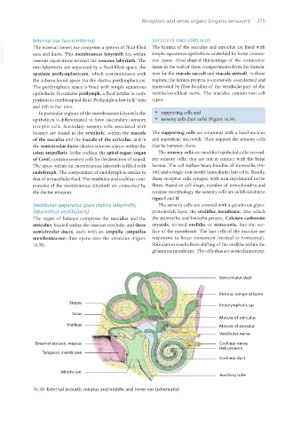
Vestibular apparatus (pars statica labyrinthi, The sensory cells are covered with a gelatinous glyco-
labyrinthus vestibularis) protein-rich layer, the otolithic membrane, into which
The organ of balance comprises the sacculus and the the stereocilia and kinocilia project. Calcium carbonate
utriculus, located within the osseous vestibule, and three crystals, termed otoliths or statoconia, line the sur-
semicircular ducts, each with an ampulla (ampullae face of the membrane. The hair cells of the maculae are
membranaceae) that opens into the utriculus (Figure responsive to linear movement (vertical or horizontal).
16.38). Stimulation results from shifting of the otoliths within the
gelatinous membrane. The cells thus act as mechanorecep-
16.38 External acoustic meatus and middle and inner ear (schematic).
Vet Histology.indb 375 16/07/2019 15:08